Explore the World of Digital Microscopy with EVOS
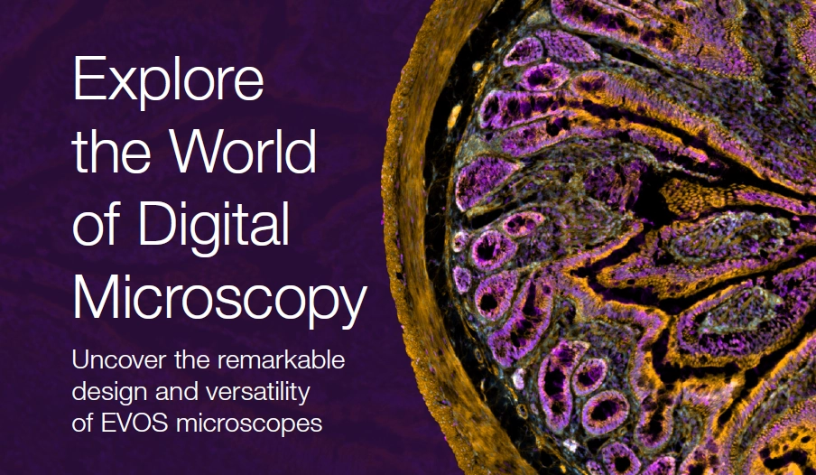
Discover how EVOS microscopes are transforming imaging in modern life sciences
What You'll Learn:
- Common limitations of traditional microscopes—and how EVOS overcomes them
- Side-by-side comparisons of EVOS M3000, M5000, and M7000 imaging systems
- Key onboard features: real-time confluency, 2D deconvolution, and stage tracking
- How researchers worldwide are using EVOS systems to publish results faster
Ideal for:
Cell and tissue imaging - Live cell imaging analysis
- Core facilities and individual research labs
Real-time image and video capture
From entry-level to advanced imaging, EVOS microscopes offer intuitive design, high-quality optics, and versatile application support—all in a compact, easy-to-use system.

DOWNLOAD THIS INFOGRAPHIC
Become a member and enjoy exclusive benefits
Create an account now for exclusive benefits, personalized recommendations, and seamless order tracking. Elevate your lab experience today!