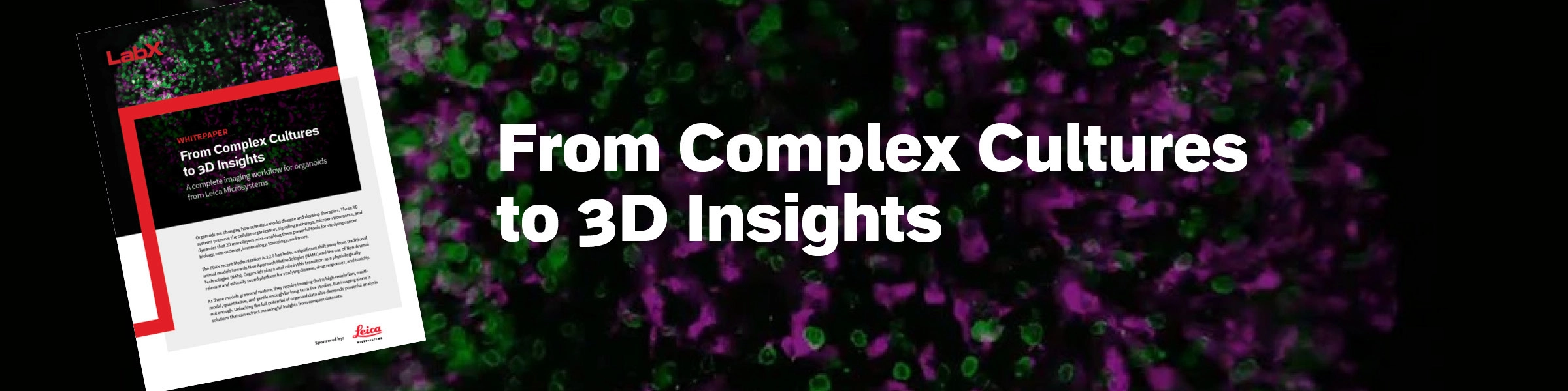
From Complex Cultures to 3D Insights
Exploring a Streamlined Workflow for Imaging Organoids from Leica Microsystems
Organoids are transforming how researchers model disease and develop therapies. As physiologically relevant 3D systems, they preserve the cellular organization, signaling pathways, and microenvironments that 2D cultures cannot replicate, and are increasingly being used as part of New Approach Methodologies (NAMs).
Extracting meaningful biological insights from these intricate and challenging models requires not only high-resolution, gentle long-term imaging, but also AI-based analysis for even deeper understanding.
This whitepaper explores a workflow from Leica Microsystems to capture, analyze, and understand complex 3D biology with greater clarity and less complexity.
- Initial organoid growth checks: Ensure readiness with the intuitive Mateo TL and FL Digital Microscopes
- Rapid pre-screening: Triage organoids quickly with THUNDER Imager Cell Spinning Disk or the Mica Imaging Microhub
- High-resolution multi-dimensional imaging: Capture precise 3D structures using the STELLARIS confocal platform
- Long-term 3D live studies: Track growth and response dynamics over time with the Viventis Dual-View Light-Sheet Microscope
- AI-powered image analysis: Transform complex datasets into quantifiable insights using Aivia AI Image Analysis Software
Together, these tools create a streamlined imaging ecosystem—from initial culture checks to analysis—enabling reproducible, physiologically relevant results in areas such as cancer, neuroscience, and regenerative medicine research.

DOWNLOAD THIS WHITEPAPER TO LEARN MORE
Become a member and enjoy exclusive benefits
Create an account now for exclusive benefits, personalized recommendations, and seamless order tracking. Elevate your lab experience today!