
Optimizing 3D Cell Culture Assays by Leveraging Kinetic Imaging and Endpoint Viability Analysis
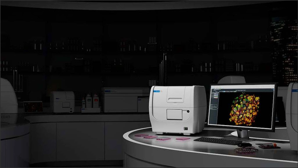
Image- and luminescence-based viability quantification for scalable treatment-response characterization of 3D cell culture models
Key challenges in 3D screening—such as model quality, focus, and tracking treatment-response trajectories—limit the ability to support new approach methodology (NAM)-relevant drug discovery.
This application note details an optimized, high-throughput workflow for characterizing three-dimensional (3D) cell culture models (spheroids) by combining automated kinetic imaging with orthogonal endpoint viability analysis using the Agilent BioTek Cytation 9 cell imaging multimode reader.
Takeaways
Download the whitepaper to learn more.

DOWNLOAD THIS APPLICATION NOTE
Become a member and enjoy exclusive benefits
Create an account now for exclusive benefits, personalized recommendations, and seamless order tracking. Elevate your lab experience today!