
Combine SEM Imaging and Elemental Analysis for Materials Characterization
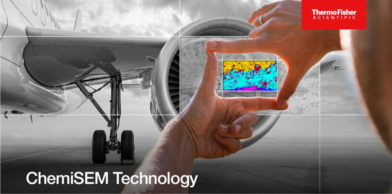
Reveal defects and contaminants that grayscale SEM images can miss.
Materials researchers increasingly use
scanning electron microscopy (SEM) to study composition as well as structure.
When imaging and elemental analysis run in separate programs, analysts juggle
two interfaces to investigate one sample. This guide from Thermo Fisher Scientific
shows how ChemiSEM Technology runs SEM and energy-dispersive X-ray spectroscopy
(EDS) from one workspace. What You'll Learn Download the guide to judge whether
ChemiSEM Technology on Axia or Apreo 2 fits the materials your lab
characterizes.

DOWNLOAD THIS GUIDE TO LEARN MORE
Become a member and enjoy exclusive benefits
Create an account now for exclusive benefits, personalized recommendations, and seamless order tracking. Elevate your lab experience today!