Advancing Live-Cell and Time-Lapse Imaging with Digital Microscopy
Transforming live-cell and time-lapse imaging from labor-intensive manual methods into scalable, automated high-throughput workflows
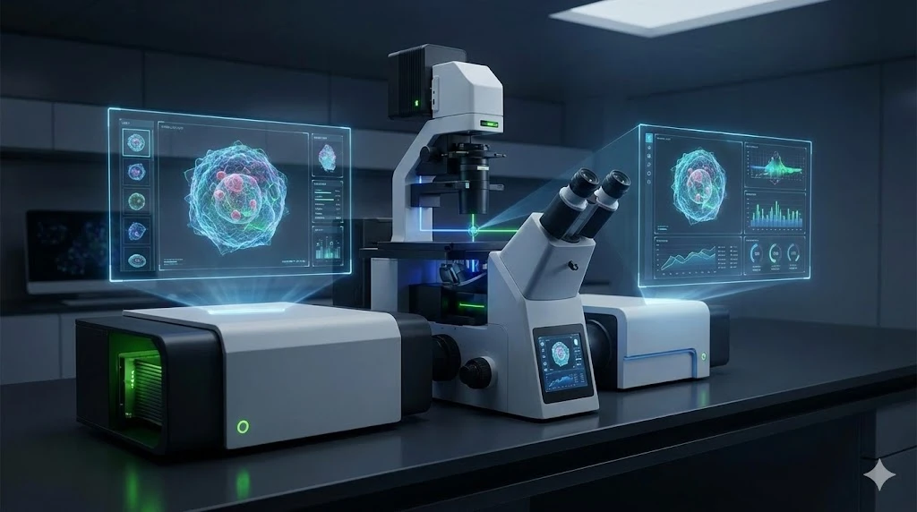
Advancements in automated digital fluorescence microscopy are significantly enhancing live-cell and time-lapse imaging, two essential techniques in modern cell biology and biomedical research. These innovations are not only streamlining workflows but also expanding experimental capabilities, allowing scientists to observe dynamic cellular processes with unprecedented precision, consistency, and scale. Here are several aspects of technology development that are leading the way:
1. Maintaining Optimal Imaging Conditions Over Time
Live-cell imaging requires precise environmental control to keep cells healthy and responsive throughout the duration of an experiment. New automated digital systems include:
- Integrated incubation chambers to maintain temperature, humidity, and CO₂ levels
- Automated focus stabilization that compensates for thermal drift and sample movement
- Adaptive illumination to minimize phototoxicity and photobleaching over long-term imaging
These features are designed to ensure consistent image quality over hours—or even days—of continuous monitoring, making time-lapse studies more reliable.
2. Improved Temporal Resolution and Scheduling
Digital fluorescence systems allow researchers to program complex, high-frequency imaging schedules that would be impractical to perform manually. Automated capabilities include:
- Precise interval timing for high-resolution kinetic data
- Multi-position scanning to monitor multiple regions of interest across wells or slides
- Dynamic feedback loops, where imaging parameters can adjust in response to biological events (e.g., a change in fluorescence intensity)
This enables researchers to capture fast cellular events like mitosis, vesicle trafficking, or calcium signaling with consistent temporal accuracy.
3. Multiplexed and Multi-Channel Imaging
In time-lapse experiments, it’s often critical to observe multiple fluorophores simultaneously or sequentially. Automated digital microscopes can:
- Switch fluorescence channels rapidly and precisely using motorized filter wheels or tunable light sources
- Synchronize illumination and image capture to avoid bleed-through and cross-excitation
- Correct for channel misalignment and drift over time using built-in software algorithms
These capabilities support complex, multi-dimensional experiments such as tracking protein translocation, monitoring gene expression dynamics, or following intracellular signaling cascades.
4. Scalability and High-Content Time-Lapse Studies
With motorized stages and multi-well plate compatibility, automated systems enable time-lapse imaging of hundreds of samples simultaneously. This opens doors to:
- Drug screening over time in a live-cell context
- Stem cell differentiation monitoring across varying conditions
- Phenotypic profiling in genetically modified cell lines
Coupled with automated image analysis, researchers can quickly identify patterns or outliers across vast datasets.
5. Enhanced Data Integrity and Analysis
Modern systems support direct integration with image analysis platforms and data management tools, offering:
- Real-time tracking of cell morphology, motility, division, and apoptosis
- Automated segmentation and quantification
- Cloud-based storage and sharing for long-term studies
This shift from image capture to data-driven insights significantly boosts experimental reproducibility and interpretation.
Conclusion
Automated digital fluorescence microscopy is transforming live-cell and time-lapse imaging from labor-intensive niche methods into scalable, high-throughput workflows. By combining environmental control, temporal precision, and intelligent automation, these systems empower researchers to ask—and answer—complex biological questions with confidence.
Download the whitepaper to learn about new technologies such as the EVOS imaging systems defining new frontiers in the digital microscopy world.

