Electron Diffraction of Nanocrystals — Going beyond the Limits of X-Ray Diffraction with the Rigaku XtaLAB Synergy-ED

Written By: LabX.com
Jul 06, 2022
Introduction
Electron Diffraction, sometimes also known as 3DED/microED (3-Dimensional Electron Diffraction or micro Electron Diffraction), is a revolutionary analytical technique for the advancement of structural science. Previously, crystallographers relied on single crystal X-ray diffraction (SC-XRD), which restricted them to samples of the order of 1 µm. With the advent of electron diffraction, the limitations of SC-XRD have been overcome and the structural determination of nanocrystals in the realms of tens of nanometers is now possible.
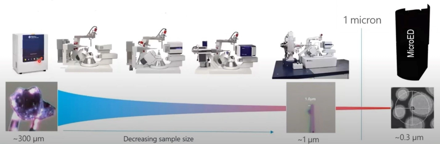
Figure 1. Representation of analytical instruments and technologies vs. crystal size.
Prior to electron diffraction becoming available, synthetic chemists were forced to use NMR to try to postulate the 3D structure of new materials that only formed nanocrystals. For complex molecules, NMR is extremely challenging to interpret. As a result, researchers looked for alternatives. Early works published by Kolb et al. (2007) and Shi et. al. (2013) paved the way for the development of this new technique.
Recognizing the potential of electron diffraction for the crystallography community, Rigaku and JEOL embarked on a partnership to develop the world’s first commercially available electron diffraction system in 2020. The following year, the collaboration culminated in the XtaLAB Synergy-ED, a dedicated electron diffractometer. The XtaLAB Synergy-ED fully integrates expertise from both companies, i.e.:
- Rigaku’s high-speed, high-sensitivity detector (HyPix-ED) and high-powered crystallographic software
- JEOL’s expertise in generation and control of stable electron beams
The system incorporates a seamless workflow from data collection to structure determination of three-dimensional molecular structures using CrysAlisPro-ED, an extension of Rigaku’s proven CrysAlisPro structural determination software for SC-XRD.
Electron diffraction differs from SC-XRD in that it uses electrons as the source rather than X-rays. This enables crystallographers to examine crystals smaller than the ~1 µm limit of SC-XRD. Due to the fact that electrons interact with matter to a much greater extent compared to X-rays, smaller crystals—of the order of tens of nanometers —are ideally suited to electron diffraction. This makes it a highly complementary technique to SC-XRD.
Challenges for Electron Diffraction
The challenges for electron diffraction are different to those of SC-XRD
- Absorption – Small crystals are often better, as even small crystals can completely stop the electron beam
- Dynamical Diffraction – Electron beams are diffracted multiple times as they traverse through the crystal. Therefore, the thicker the crystal, the greater the dynamical effect, which can be problematic for resolving and refining crystal structures
- Sample decay – Exposing crystals to high-energy electron beams result in decay. This can be overcome by using low temperature data collection and/or a carefully selected beam intensity and scan speed
Electron Diffraction Systems
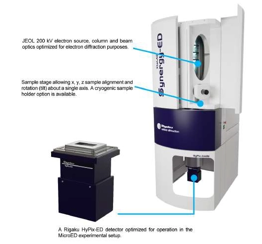
Figure 2. Rigaku XtaLAB Synergy-ED, world's forst turnkey electron diffractometer for determination of crystallographic structure of nanocrystals.
Rigaku’s XtaLAB Synergy-ED consists of a vertical vacuum column that ensures the sample stays in the beam throughout the scan. The column incorporates a JEOL 200 kV electron source and optical system.
Samples are mounted on a stage that allows x, y, z and tilt adjustment for precise sample positioning. A cryo-transfer sample holder is also available for sensitive samples.
The XtaLAB Synergy-ED uses a HyPix-ED detector. This is similar to other HyPix detectors used in SC-XRD in that it is event-driven, meaning it:
- Counts each diffracted electron
- Does not produce any dark noise or readout noise
- Offers high frame rates
- Produces high-quality data
The system is controlled using the same CrysAlisPro software package as Rigaku’s SC-XRD systems which means anyone familiar with those systems will be able to perform electron diffraction experiments almost immediately. The only difference is the sample visualizing and centering functions which are self-explanatory and intuitive. The analytical workflow is identical.
As with X-ray diffraction experiments, CrysAlisPro performs data acquisition and data processing concurrently. AutoChem, which is fully integrated into CrysAlisPro, performs fast, fully automated structure solution and refinement during data collection, thereby optimizing your productivity.
Due to the mounting approach and limited sample access in electron diffraction experiments, it is often necessary to merge data from multiple grains to generate a complete dataset. CrysAlisPro makes this a simple operation, also applying all corrections and scaling to produce the final hkl reflection file.
Sample Preparation
Preparing samples for electron diffraction experiments is relatively straight forward, involving:
- Grinding samples
- Attaching to TEM grids
- Loading grids into the XtaLAB Synergy-ED
Results
Below are examples of structures that have been determined using electron diffraction, which typically allows you to see hydrogen positions in the different maps. All samples were examined using the XtaLAB Synergy-ED using the CrysAlisPro software. Structures were refined automatically by AutoChem or manually using Olex2.
Structures were refined using SFAC instructions suitable for electron diffraction. In some instances, single grains provided sufficient completeness for structural solution. In other cases, structures were resolved by merging datasets from multiple grains.
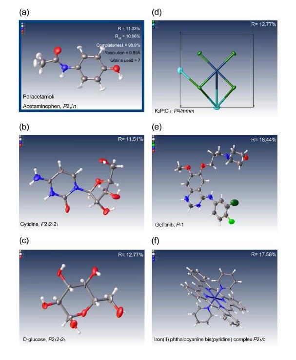
Figure 3. First examples of the first attempts at solving structures with data collected on the XtaLAB Synergy-ED including pharamaceutical compounds, iroganic materials and coplexes ranging from triclinic to tetragonal symmetry: (a) paracetemol (acetaminophen), (b) cytinide, (c) glucose, (d) potassium tetrachloroplatinate, (e) gefitnib and (f) iron(II) phthalocyanine bis(pyridine) complex.
The first published structures using data from an XtaLAB Synergy-ED system were reported by researchers from the University of Birmingham and the University of Nottingham lead by Prof. Neil Champness in the journal Nature Communications (January 2022).
The research team successfully determined the structure of the difficult rotaxane crystals using electron diffraction. This followed unsuccessful attempts to resolve the structure using X-Rays on a synchrotron beamline.
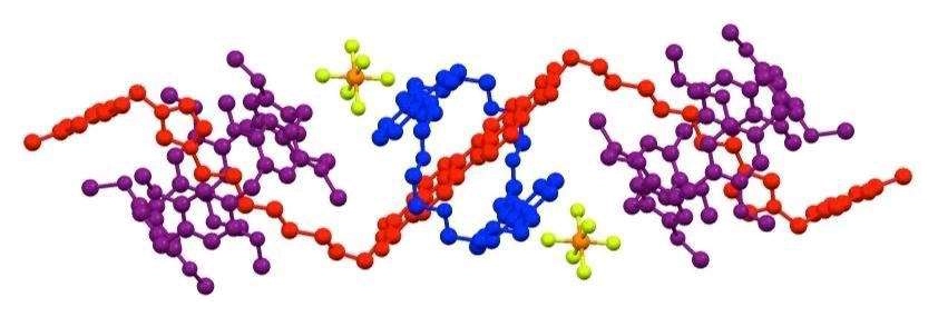
Figure 4. Rotaxane structure determined using the Rigaku XtaLAB Synergy-ED electron diffractometer.
“We are experienced X-ray crystallographers and use our own Rigaku XtaLAB Synergy-S X-ray diffractometer as well as synchrotron X-ray sources. For the compound in this study, we collected X-ray data, but it wasn't of a sufficient quality to determine a reliable structure of the molecule and then Rigaku stepped in with their new XtaLAB Synergy-ED. Much to our surprise the electron diffraction experiment gave better quality data for our crystals than even the synchrotron X-ray. We were very pleased that electron diffraction gave such good data for such a complex molecule, and this allowed a much greater appreciation of the chemistry that we are studying”, said prof. Champness.
Applications
Electron diffraction has already been used to help elucidate the structure of the Coronavirus.
This technique also has the potential in fields such as:
- Catalysis
- Energy storage and alternative energies
- Aerosol research
- CO2 reduction
- Biomineralization
- Pharmaceuticals
- Medical technology
- Mineralogy
- Archaeology
Electron Diffraction Offered as a Service
In a recent development, Rigaku are also now offering access to this state-of-the-art technology for crystallography projects in pharmaceuticals, organic and inorganic chemistry, materials science and even mineralogy as a service. Researchers from industry and academia will now be able utilize the world’s first turnkey electron diffractometer, the Rigaku XtaLAB Synergy-ED, without the expense of having to buy one. The new program provides access to instrumentation and software–as well as in-house expertise–to assist with your projects.
Conclusion
Direct structural determination of nanocrystals and samples too small for conventional X-ray diffraction studies is now possible using electron diffraction. For the first time, a totally dedicated electron diffractometer is commercially available in the XtaLAB Synergy-ED, which is able to produce structural refinements from previously unmeasurable samples. This turnkey system is the ideal complement to existing single crystal diffractometers and, due to its compact design, can be easily accommodated into existing X-ray labs.
Driven by the CrysAlisPro software interface, which is familiar to many crystallographers, you will be able to perform MicroED experiments with almost no training, with the potential to rapidly generate publications using this emerging analytical technique.
References
- Kolb et al, (2007), Towards automated diffraction tomography: Part I—Data acquisition, Ultramicroscopy, 107, (6-7), 507-513
- Shi et al. (2013), Three-dimensional electron crystallography of protein microcrystals, eLife
- Pearce et al. (2022), Selective photoinduced charge separation in perylenediimide-pillar[5]arene rotaxanes, Nature
This editorial was written by Rigaku and published in collaboration with LabX.
For more information visit Rigaku.com

Dec 24, 2025
Featured, Popular Products, Technical Insight
Emerging Mass Spectrometry Technologies to Improve Intraoperative Diagnostics
Advances in mass spectrometry may reshape the landscape of surgical oncology, improving diagnostic accuracy and potentially impacting post-operative outcomes

Dec 23, 2025
Technical Insight
The Best Microplate Washers/Dispensers of 2026
Discover the top microplate washers/dispensers for 2026. Compare the latest models from Agilent, BlueCatBio, and Thermo Fisher to optimize your lab's throughput and precision today.
Dec 15, 2025
Featured, Popular Products
Automated 3D Cell Culture Workflows: Reshaping Disease Research and Drug Development
As 3D cell systems move from exploratory research into routine use across drug development and translational science, automation is becoming essential.
Dec 11, 2025
Technical Insight
The Best FPLC Systems of 2026
Discover the top FPLC systems of 2026 for protein purification. This guide reviews price, specs, and performance for leading lab equipment models.
Dec 11, 2025
Technical Insight
The Best Fermenters/Bioreactors of 2026
Explore the top fermenters/bioreactors of 2026 for research and production. Discover high-throughput, single-use, and budget-friendly models to optimize your lab's workflow.
Dec 11, 2025
The Best ELISA Kits and Immunoassay Systems of 2026
Discover the top-rated ELISA kits and immunoassay systems of 2026. We review high-throughput analyzers and budget-friendly models for modern labs.
Dec 10, 2025
Featured, Popular Products
How Microplate Dispensers Enable Modern Research and Discovery
From drug discovery to genomics, almost every lab today depends on microplate dispensers.
Dec 08, 2025
Technical Insight
The Best Colorimeters of 2026
Discover the top-rated colorimeters of 2026 for laboratory professionals. We review the best new models for accuracy, speed, and chemical analysis.
Dec 08, 2025
Technical Insight
The Best DNA Sequencers of 2026
Discover the top-performing DNA sequencers of 2026. This guide reviews the latest high-throughput, benchtop, and long-read models for modern laboratories.
Dec 08, 2025
Technical Insight
The Best Biosafety Cabinets and Laminar Flow Hoods of 2026
Discover the top biosafety cabinets and laminar flow hoods of 2026. This guide reviews safety, efficiency, and features for optimal laboratory protection.