Mastering Flow Cytometry: Principles, Applications, and Top Instruments

Written By: Craig Bradley
Mar 07, 2018
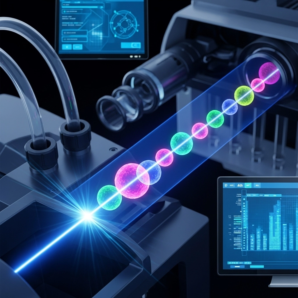
ImageFX (2025) Flow cytometry stands as a cornerstone in modern biological and medical research, offering unparalleled insights into cellular characteristics. This sophisticated biophysical technique empowers scientists to rapidly analyze and sort cells based on their unique properties, primarily through the emission of fluorescence. From fundamental research to clinical diagnostics, flow cytometry has transformed our understanding of cellular populations, enabling high-throughput, multi-parameter analysis of thousands of particles per second. Dive in to discover how this essential technology works, its myriad applications, and the cutting-edge instruments driving innovation in laboratories worldwide. At its heart, a flow cytometer is an intricate system designed for precision cell analysis. It meticulously guides cells, suspended in a fluid stream, through a focused light beam and past a detector. Unlike traditional microscopy, which captures static images, flow cytometry provides dynamic, cell-by-cell quantitative data, making it exceptionally powerful for real-time monitoring and high-throughput studies. A typical flow cytometer comprises five essential compartments working in harmony: Flow Cell: This critical component uses a "sheath fluid" to hydrodynamically focus and align cells, ensuring they pass through the laser interrogation point in a single file. This single-file alignment is crucial for accurate individual cell analysis. Measurement System: Equipped with various laser sources—ranging from high-power water-cooled to compact air-cooled and diode lasers—this system excites fluorophores attached to or within the cells. The choice of laser depends on the specific fluorophores and application requirements. Detector System: As excited cells pass through the laser, they emit light signals. Detectors capture these signals, including forward-scatter (FSC) for cell size, side-scatter (SSC) for internal complexity (granularity), and various fluorescence signals specific to tagged cellular components. Amplification System: The raw light signals from the detectors are weak. This system amplifies these analog signals and converts them into digital data, preparing them for computational processing. Data Analysis System: Sophisticated software processes the digital signals, generating plots and statistics that allow researchers to identify, quantify, and characterize distinct cell populations based on their light scattering and fluorescence properties. The sensitivity and real-time capabilities of flow cytometry, particularly in fluorescence quantification, often surpass traditional microscopy methods, which are limited to collecting in-focus signals from individual measurements. Modern flow cytometers are incredibly advanced, featuring multiple lasers (sometimes up to ten) and numerous fluorescence detectors (up to 30), enabling highly complex multi-color assays. This technological evolution, coupled with an ever-expanding catalog of fluorescent probes and antibodies, has cemented flow cytometry's indispensable role in both research and clinical laboratories. A highly specialized and transformative application of flow cytometry is Fluorescence Activated Cell Sorting (FACS). This technique takes the analytical power of flow cytometry a step further by enabling the physical separation of mixed cell populations into distinct "bins" based on their unique fluorescence characteristics. The process of FACS involves an elaborate, multi-step mechanism: Cell Alignment and Detection: Similar to standard flow cytometry, cells are aligned in a fluid stream and pass through a laser, where their fluorescence and light scatter properties are detected. Droplet Formation: After detection, the fluid stream containing the cells is vibrated at a high frequency, causing it to break into discrete, uniform droplets. Crucially, each droplet ideally contains only one cell. Electrostatic Deflection: Based on the detected fluorescence signals, an electric charge is applied to droplets containing cells of interest. These charged droplets then pass through an electrostatic deflection system, which uses an electric field to divert them into separate collection tubes or wells. Uncharged droplets (containing unwanted cells or no cells) are discarded. FACS is a powerful tool for isolating pure populations of cells for further study, culture, or therapeutic applications. It's important to note that "FACS" is an acronym that was trademarked by Becton Dickinson, a leading manufacturer in the field. For more in-depth methods and troubleshooting guides, explore our full guide on flow cytometry fundamentals. The versatility of flow cytometry is one of its most compelling attributes. Its ability to analyze a wide array of cell types, combined with an incredibly diverse collection of fluorescence-emitting antibodies and probes, makes it invaluable across numerous scientific disciplines. Key applications include: Surface Staining (Direct & Indirect): Direct Staining: Utilizes fluorophore-conjugated primary antibodies to directly detect surface-exposed epitopes, antibodies, or proteins on cells. This is particularly effective for analyzing biofluids like blood to characterize immune cells and factors. Indirect Staining: Employs a fluorophore-conjugated secondary antibody to detect a primary antibody that has bound to the target. This method is useful when a conjugated primary antibody isn't available or when signal amplification is required. Intracellular Staining: Enables the detection of intracellular targets by staining fixed or permeabilized cells. Both direct and indirect staining techniques can be adapted with appropriate sample preparation. DNA Content Analysis: Crucial for cell cycle research, allowing scientists to quantify DNA content and assess cell proliferation, apoptosis, and ploidy. Cell Viability and Apoptosis Assays: Identifying live, dead, or apoptotic cells using specific fluorescent probes. Phenotyping and Immunophenotyping: Characterizing cell types and subtypes based on the expression of specific surface or intracellular markers, especially critical in immunology and hematology. Functional Assays: Measuring cellular functions such as calcium flux, cytokine production, and oxidative burst. The broad utility of flow cytometry methods and research applications underscores its significant value across fields ranging from immunology and oncology to microbiology and stem cell research. The continuous evolution of flow cytometry technology has led to the development of highly sophisticated instruments, each offering unique advantages to suit diverse research needs. Here are some prominent examples of modern flow cytometers: Beckman Coulter Gallios Flow Cytometer Research SystemThe Gallios system is renowned for its analytical excellence, combining high sensitivity, resolution, and dynamic range with rapid data collection. Superior Detection: Offers advanced optical design and sensitivity optimized for multi-color assays. Streamlined Workflow: Features user-friendly software and automation capabilities to simplify complex multi-color flow cytometry assays. Scalable Formats: Available in four scalable configurations, providing versatility in laser and fluorescence detector applications. It can accommodate up to four solid-state lasers and acquire up to six fluorescence signals (or more depending on concurrent reading needs). Enhanced Productivity: Designed to significantly improve user workflow and experimental throughput. Cytek Aurora Flow Cytometer
Cytek's Aurora series sets new benchmarks for flexibility and sensitivity, excelling at resolving rare or dim cell populations, all while maintaining affordability. Full Spectrum Capabilities: Utilizes an innovative optical design that captures the full emission spectrum of fluorophores, enabling the use of a wide array of new fluorophore combinations without system reconfiguration. High Parameter Analysis: Features three lasers, three scattering channels, and an impressive capacity for up to 48 fluorescence channels, making it suitable for a vast range of simple to highly complex applications. Exceptional Performance: State-of-the-art low-noise electronics and flat-top beam profiles, combined with a uniquely designed fluidic system, ensure outstanding sensitivity, resolution, and performance even at high sample flow rates. Intuitive Software: The SpectroFlo™ software provides an intuitive workflow, from quality control to data analysis, with technology-enabling tools that simplify running any application. Sysmex CyFlow® Cube 8/Cube 8 Max Flow Cytometer
The CyFlow Cube 8 is a compact yet powerful flow cytometry analyzer, offering modular configurations that can be customized for specific research demands. Compact & Integrated Design: Features a small footprint with a built-in Windows PC, 19" LCD screen (with second screen support), software-controlled pressure regulators, and integrated sheath/waste containers. Modular Versatility: Provides upgrade options for optical parameters and fluorescence channels, with a wide range of nine excitation wavelengths (355 - 785 nm) for additional laser light sources. Automation Ready: Compatible with optional CyFlow Sorter and CyFlow Robby 8 Autoloading Station for automated processing of well plates and sample tubes. Enhanced Max Series: The CyFlow Cube 6 Max and Cube 8 Max models include powerful Blue 488 nm and Red 640 nm lasers, enhanced optical filters, and the upgraded CyFlow software for easy-to-use yet powerful results. Flow cytometry is an indispensable technology that continues to drive discovery and innovation across the life sciences. Its ability to provide rapid, multi-parameter analysis and precise cell sorting has made it a foundational tool for understanding cellular biology, diagnosing diseases, and developing new therapies. As the technology advances, offering greater sensitivity, higher throughput, and more intuitive interfaces, its impact will only continue to grow. Explore the latest flow cytometry solutions and discover how LabX can support your research needs. Visit LabX today! What is the primary purpose of flow cytometry?
Flow cytometry is primarily used for the rapid, multi-parameter analysis and sorting of individual cells or particles in a fluid stream, based on their light scattering and fluorescence properties. How does Fluorescence Activated Cell Sorting (FACS) differ from standard flow cytometry?
FACS is a specialized type of flow cytometry that not only analyzes cells but also physically separates them into distinct populations based on their detected fluorescence characteristics. What are common applications of flow cytometry in research?
Common applications include immunophenotyping, cell cycle analysis, apoptosis detection, intracellular protein analysis, and the study of cell viability and function in various biological and medical research fields. What are the key components of a flow cytometer?
A flow cytometer typically consists of a fluidics system (flow cell, sheath fluid), an optical system (lasers, lenses, filters), a detector system (photomultipliers), an amplification system, and a data analysis system.
Understanding Flow Cytometry: Core Principles and Mechanism
Fluorescence Activated Cell Sorting (FACS): Precision Cell Separation
Diverse Applications of Flow Cytometry in Research & Clinical Settings
Leading Flow Cytometry Instruments: Features & Innovations
Final Thoughts on Flow Cytometry
Frequently Asked Questions (FAQ)

Jan 09, 2026
Product News
BRANDTECH Scientific Introduces the Transferpette® pro Micropipette: A New Twist on Comfort and Control
The latest addition to its trusted line of precision liquid handling instruments, engineered for comfort, control, and repeatable accuracy.

Jan 06, 2026
Featured, Technical Insight
New Research Frontiers Unlocked by Next-Generation Flow Cytometry
Next-generation flow cytometry systems using acoustic focusing are opening entirely new areas of research that were previously impractical or inaccessible with conventional flow cytometers.

Jan 05, 2026
Buying Guides
The Bottom Line: How Smart Microcentrifuge Tube Choices Impact Lab Efficiency and Cost
Innovative microcentrifuge tube solutions are more than just lab consumables—they’re strategic tools that can reduce costs, improve outcomes, and support long-term sustainability goals.

Jan 05, 2026
Buying Guides
Future-Proofing Your Lab with Innovative Microcentrifuge Tube Solutions
As laboratories evolve to meet the growing demands of modern science, even the most basic tools—like microcentrifuge tubes—are being reimagined.

Jan 05, 2026
Buying Guides
Microcentrifuge Tube Innovations: Driving Sustainable Change
Integrating next-generation consumables into standard workflows can lead to a more sustainable and responsible research environment.

Jan 05, 2026
Buying Guides
Single Use Plastics: How to Manage these Major Contributors to Lab Waste
Reducing lab waste isn’t just a sustainability issue—it’s a financial and operational one, too.

Jan 05, 2026
Buying Guides
Common Microcentrifuge Tube Challenges in Sensitive Molecular Workflows
Key factors that can impact the performance of microcentrifuge tubes for PCR, qPCR, and NGS and other sensitive applications

Jan 05, 2026
Buying Guides
How to Research the Right Microcentrifuge Tubes for Your Application
Selecting the right microcentrifuge tubes starts with understanding the specific demands of your application.

Jan 05, 2026
Technical Insight
The Best Confocal Microscopes of 2026
Discover the best confocal microscopes of 2026 for advanced imaging. Compare top models from Zeiss, Evident, and Nikon for speed, resolution, and budget.

Dec 24, 2025
Featured, Popular Products, Technical Insight
Emerging Mass Spectrometry Technologies to Improve Intraoperative Diagnostics
Advances in mass spectrometry may soon reshape surgery, improving diagnostic accuracy and impacting post-operative outcomes