Innovations in Cell Imaging and Multi-mode Detection Devices
Multi-mode functionality is a growing trend particularly among microplate technologies. Whether absorbance detection, fluorescence, or brightfield cell imaging -- when paired with traditional microplate processing, the advantages of this functionality are many fold.
The microplate sample format allows for streamlined sample or cell culture prep with batch reproducibility. This format is well suited for interfacing with multiple processing and detection devices, thus increasing throughput and efficiency. Advanced instrument designers such as Biotek, have brought several of these features and capabilities together under one lid. The Cytation line of instruments are prime examples of this innovation and efficiency.
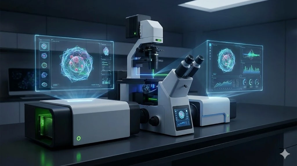
The Cytation 5 Cell Imaging Multi-mode Reader combines traditional widefield microscopy imaging with UV-Vis absorbance detection. This technology incorporates a patented design that produces both qualitative cellular information as well as quantitative well-based data, without the costs and complexity of more typical digital microscopy systems. The optics and detection components are physically and operationally separate, ensuring optimized and straightforward performance. Coupled with the Gen5 software, image capture, processing, and analysis can be performed via a seamless workflow. Biotek has coined the term "augmented microscopy" to indicate the combination of all of these features together in one instrument platform.
This concept has been taken one step further with the recent release of the Cytation 1 Cell Imaging Multi-mode Reader. This instrument combines fluorescence and high contrast brightfield imaging with conventional multi-mode detection. The detection module includes high-sensitivity filter-based fluorescence and a monochromator system for UV Vis absorbance. The Gen5 software creates a streamlined workflow that is both powerful and yet relatively simple to use. A couple specifications:
- Microscopy imaging objectives range from 1.25X to 60X, permitting visualization of large regions of interest of intracellular features. More than 15 filter LED cubes are available, expanding imaging applications.
- Automation capabilities include automated XY stage movement, focus, auto exposure and image capture, and LED illumination intensity.
- Cell and sample supportive features including: 4-zone temperature control to 45 °C with condensation control, Co2-O2 control, shaking control, angled injectors, and others. These features facilitate live cell imaging and longer-term kinetic experiments.
- Detection modes include: UV-Vis absorbance, Fluorescence intensity, Luminescence, Fluorescence polarization, and Time resolved fluorescence.
- Read modes include: Endpoint, Kinetic, Spectral Scanning, Well area scanning.
- Microplate capability includes: 6- to 384-well plates with monochromator, 6- to 1536-well with filters, and 6- to 1536 well plates for imaging applications.
- The instrument is compatible with Biotek Biostack and 3rd party automation, as well as Biospa Automated Incubator devices.
The full range of specifications can be found here.
The real power in this technology comes from the ability to capture rich cellular information and accurate quantitative measurements in a single experiment. Coupling these analyses removes the error associated with separate sample preparation and detection processes, and provides higher resolution regarding cell localization and kinetic mechanisms. The range of applications include:
Live Cell Imaging – Physiological processes have both spatial and temporal aspects. In order to accurately capture events such as cell signalling by second messengers, cytokines, or other molecules it is often mandatory to measure changes on the millisecond time-scale. A detection system with these capabilities is therefore necessary. Quantitative processes such as cell growth or motility also require high resolution imaging and detection capabilities. Furthermore, cell viability may require the right environmental factors, such as specific temperature, gas concentrations, and humidity – conditions delivered from the proper instrumentation.
Immunofluorescence – Cell labelling with antibodies and imaging by fluorescent probe detection is a popular method used in many fields. Requirements include high resolution microscopy and accurate fluorescent channeling to ensure the highest value results. The Cytation 1 provides high magnification and 19 different fluorescence channels to choose an emission spectrum spanning deep blue to red. The optional automated workflow for fixing, permeabilizing, and staining cells provides a further level of performance.
Phenotypic Assays – Complex physiological processes may involve cell signalling, morphological changes, dynamic cell death or proliferation, and other events. The Cytation 1 allows multiple processes such as these to be followed in real time, through the combination of Brightfield imaging, Immunofluorescence, Live (and Dead) cel counting, etc. The combination of functions makes this platform powerful for complex applications such as neuroscience and developmental research.
There are other capabilities as well, including multi-mode absorbance reading and label-free cell counting, which add to the versatility of the platform.
The Cytation 1 is well suited for Neuroscience research owing to its ability to image cells, monitor processes, and quantitate signals, simultaneously on the time-scale required. It is the latest innovation from Biotek and sure to be heralded as a star performer.
