Innovative Digital Solutions to the Limitations of Manual Fluorescence Microscopy
How digital systems are moving the field forward to enable reproducible, scalable, and quantitative analysis
Fluorescence microscopy remains a cornerstone across cell biology, pathology, pharmacology, and materials science. But many labs still rely on manual, observer-driven workflows built around stand-alone microscopes, hand-tuned acquisition, and post-hoc analysis. These approaches often struggle to deliver reproducible, scalable, and quantitative data at the pace modern projects demand.
This article outlines where traditional manual techniques fall short and how innovative digital microscopy systems—combining automated hardware, controlled environments, and integrated analysis—address those gaps for higher quality results with lower variability.
Where Traditional Manual Techniques Struggle
1) Reproducibility and Observer Bias
Manual exposure settings, focus, ROI selection, and thresholding decisions vary from user to user and even within the same user over time -- complicating comparisons across experiments, batches, or sites.
Impact: Higher variance, lower statistical power, and fragile conclusions that are hard to replicate.
2) Throughput and Scalability
Manually scanning multiple fields, Z-planes, or time points is slow and fatiguing. Expanding from a few images to plate-scale experiments (e.g., 24–384 wells) quickly becomes impractical.
Impact: Limited sample size, reduced experimental breadth, and longer timelines for decision-making.
3) Quantitation Limits
Manual ROI drawing and ad-hoc thresholding are time-consuming and inconsistent. Intensity drift, non-uniform illumination, and variable background make quantitative measurements difficult.
Impact: Semi-quantitative outputs that undercut robust trend detection and dose–response modeling.
4) Phototoxicity and Photobleaching
Manual oversampling and irregular exposure control increase light dose. Live-cell experiments are especially vulnerable to induced stress and bleaching that confounds biology.
Impact: Biological artifacts, signal loss over time, and unusable time-lapse sequences.
5) Environmental Instability
Temperature, CO₂, humidity, and vibration are often managed piecemeal. Even minor fluctuations during long acquisitions shift focal planes or alter cell behavior.
Impact: Focus drift, morphological changes unrelated to treatment, and inconsistent kinetic data.
6) Fragmented Data and Metadata
Images live on local drives with incomplete metadata (objective, binning, exposure, stage map, calibration). Manual notes may omit critical settings.
Impact: Weak traceability, difficult audits, and barriers to FAIR data principles (Findable, Accessible, Interoperable, Reusable).
7) Ergonomics and Training
Prolonged eyepiece work strains users, and training new operators to match expert settings is slow. Subtle acquisition choices are hard to document and transfer.
Impact: Burnout risk, uneven skill distribution, and hidden costs from rework.
What Innovative Digital Microscopy Systems Deliver
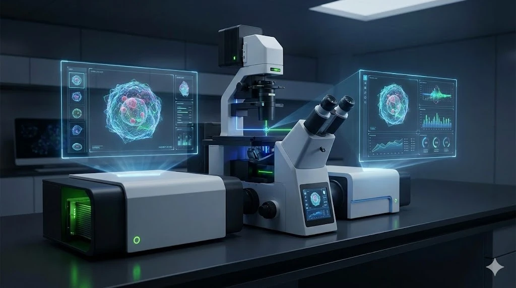
Modern digital platforms integrate motorized optics, automated stages, stable illumination, environmental control, and on-board or cloud analysis. The result is standardized, scriptable imaging that scales from a few fields to high-content screens—without sacrificing data quality.
Applications of High-Value Digital Systems
- Live-Cell and Time-Lapse Imaging: Stable environments and drift control yield clearer dynamics (division, migration, trafficking) over hours to days, enabling reliable kinetic models.
- High-Content Screening: Automated multi-well acquisition plus AI-assisted analysis scales phenotypic assays and accelerates hit triage.
- 3D and Thick Samples: Repeatable Z-stacks with deconvolution produce cleaner volumetric data for spheroids, organoids, and tissue sections.
- Co-Localization & Ratiometric Studies: Consistent illumination and calibration improve channel alignment and ratio accuracy.
- Quantitative Morphometry: Standardized segmentation enables robust statistics on cell size, shape, texture, and intensity distributions across large cohorts.
Summary
- Manual fluorescence workflows are constrained by variability, limited throughput, and fragile quantitation—especially in live-cell and longitudinal studies.
- Digital microscopy systems standardize acquisition and analysis, stabilize the environment, and embed quality control, producing reproducible, scalable, and truly quantitative data.
- Adoption doesn’t require a big-bang transition -- just an investment in the latest technology.
Download the infographic and check out the latest digital microscopy solutions designed to enable high-quality cell and tissue imaging results.


