3D Cell Imaging and Analysis: How the Landscape Is Changing

Written By: Damon Anderson, PhD
Nov 21, 2025
Reshaping expectations for 3D cell imaging and analysis and driving the development of more integrated, automated solutions.
From 2D Snapshots to 3D, Live, and Quantitative Data
Traditional fluorescence and brightfield imaging were built around thin, mostly static samples. In contrast, organoids and other 3D cultures:
- Self-organize into mini-tissues with native-like architecture and polarity
- Maintain heterogeneous cell populations and complex extracellular matrices
- Exhibit realistic gradients of oxygen, nutrients, and signaling molecules
- Produce gene expression and drug response profiles that more closely mimic in vivo biology
As a result, imaging workflows have had to expand from simple snapshots to multi-step, multi-modality processes that support:
- Routine culture checks to standardize when models are ready for experiments
- Fast pre-screening across many wells or conditions
- High-resolution volumetric imaging deep into light-scattering samples
- Long-term live imaging over hours to days
- Robust image analysis that can handle large, high-dimensional datasets
Turning 3D Visibility into Actionable Insight
3D cell imaging has moved from niche capability to a central requirement for cutting-edge biology and translational research. Vendors are moving away from isolated instruments toward end-to-end workflows that connect culture monitoring, imaging, and analysis into a coherent pipeline. As models grow more complex, the most successful labs will be those that:
- Treat imaging as a connected workflow, not a standalone step
- Prioritize gentle, high-content acquisition that preserves sample health
- Rely on AI-driven analysis to handle data scale and complexity
- Build standardized, auditable processes compatible with evolving regulatory expectations
For teams working with organoids and other advanced 3D models, now is the time to reassess whether your imaging pipeline can truly support the questions you’re asking. Download the whitepaper "From Complex Cultures to 3D Insights" to learn more about emerging solutions to the challenges of 3D model imaging.
Feb 19, 2026
Technical Insight
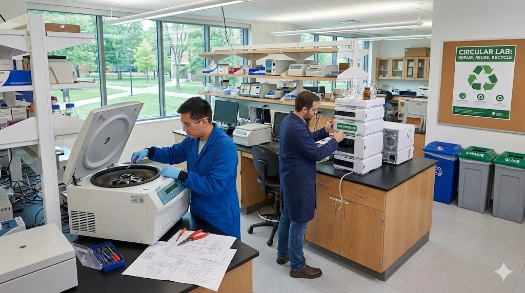
Circular Economy in Laboratory Equipment: How to Adopt Repair, Reuse, and Refurbishment Strategies
Adopting a circular economy for laboratory equipment minimizes environmental impact while maximizing instrument lifespan and optimizing laboratory budgets.
Feb 12, 2026
Product News
$5M+ in GMP Biopharma Manufacturing & QC Equipment Heads to Online Auction
A Southern California GMP manufacturing and clinical-stage QC laboratory is liquidating new and late-model equipment and unused inventory in an online auction opening February 11, 2026

Feb 12, 2026
Product News
2026 SLAS: Inspiration Catalyzing Discovery
A LabX look at the annual conference and exhibition, a key forum for networking, collaboration, and advancement of new technologies
Feb 11, 2026
Product News
Angstrom Scientific Named U.S. Distributor for Radalytica’s RadalyX Robotic X-ray and CT System
Angstrom Scientific, known for representing microscopy and materials analysis technologies across North America, adds Radalytica’s robotic X-ray and CT platform to its portfolio of analytical and imaging solutions.
Feb 10, 2026
Technical Insight
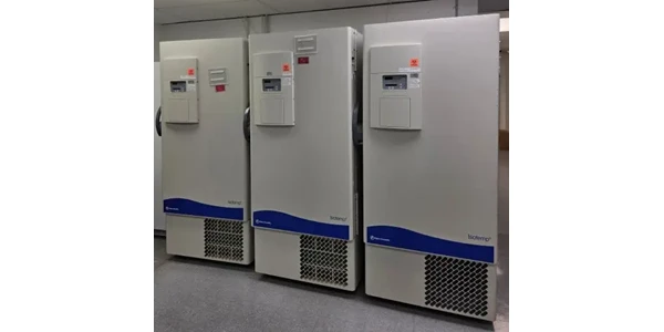
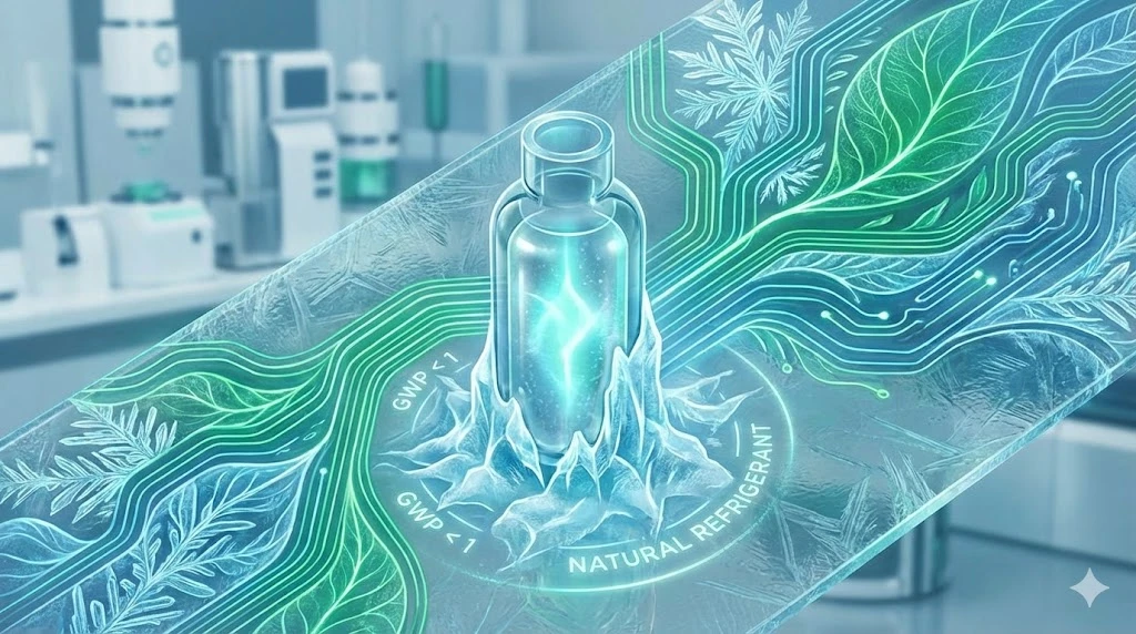
Eco-Friendly Refrigeration: Evaluating Natural Refrigerants and Low-GWP Cooling Systems
This guide explains the operational and environmental benefits of adopting natural refrigerants and low-GWP cooling systems in laboratory settings

Feb 10, 2026
Technical Insight
The Ultimate Guide to Sustainability in Laboratory Equipment and Operations
A comprehensive analysis of sustainable laboratory practices, focusing on energy-efficient equipment, waste management, and digital workflows

Jan 09, 2026
Product News
BRANDTECH Scientific Introduces the Transferpette® pro Micropipette: A New Twist on Comfort and Control
The latest addition to its trusted line of precision liquid handling instruments, engineered for comfort, control, and repeatable accuracy.

Jan 06, 2026
Featured, Technical Insight
New Research Frontiers Unlocked by Next-Generation Flow Cytometry
Next-generation flow cytometry systems using acoustic focusing are opening entirely new areas of research that were previously impractical or inaccessible with conventional flow cytometers.

Jan 05, 2026
Buying Guides
The Bottom Line: How Smart Microcentrifuge Tube Choices Impact Lab Efficiency and Cost
Innovative microcentrifuge tube solutions are more than just lab consumables—they’re strategic tools that can reduce costs, improve outcomes, and support long-term sustainability goals.

Jan 05, 2026
Buying Guides
Future-Proofing Your Lab with Innovative Microcentrifuge Tube Solutions
As laboratories evolve to meet the growing demands of modern science, even the most basic tools—like microcentrifuge tubes—are being reimagined.