LabX Highlights of the 2017 Society for Neuroscience Conference

Written By: Damon Anderson, PhD
Dec 06, 2017
"The human brain is the most complicated biological structure in the known universe. We've only just scratched the surface of how it works." These were the opening words of Francis Collins, Director of the National Institutes of Health when presenting the BRAIN Initiative, a program that was soon after launched by the president in 2013.
This overall sentiment seemed to be reflected among the masses (over 30,000) who attended the 2017 Society for Neuroscience Conference in Washington, DC November 11-15. The brain is ultimately complex and there is much to accomplish in understanding processing and function. Despite this gravity, it is clear that progress in neuroscience is being driven from all sides, at the molecular, cellular, and systems levels. Also apparent at the conference, was the myriad of new technologies and enabling solutions which are driving research and offer promise for the ever-quickening pace of discovery.
The Neuroscience conference traditionally has the scope and breadth to cover all areas of industry, from reagents, to electrophysiology, to imaging systems and well beyond. One area of focus this year - from manufacturers large and small – appeared to be high resolution imaging technologies and data analysis solutions. These technologies are attempting to visually address the range, complexity, and sheer magnitude of neuronal systems in the human brain – with an average synapse size of 4 microns, 125 trillion synapses, 100,000 miles of nerve fibers, and 86 billion neurons.
Zeiss had on display their range of instrumentation and accessories designed to suit modern neuroscience research. The company is built on 170 years of expertise and innovation, and the current instrument offerings span a wide range of applications. The Axio line of instruments were present including the Axio Examiner fixed-stage microscope, which serves as an imaging platform with space to support various electrophysiology equipment, electrodes, micromanipulators, etc. The instrument is ideal for patch clamp studies of neuronal tissue, in which voltages changes and fluorescence imaging is required.
Beyond the host of high resolution fluorescence, confocal, and multiphoton imaging platforms, the Zeiss series of scanning electron microscope solutions were on full display. The overarching theme and predominant application for these instruments are for 3-dimensional data acquisition and mapping of neuronal tissue. The field emission SEM instruments from the Sigma and GeminiSEM families interface with 3View software from Gatan, creating the ideal tools for 3-D reconstruction of neural tissue. As an alternative to serial block-face electron microscopy, the Atlas and Atlas 3D produce large area imaging of serial sections, with the option to link fluorescent markers for higher depth analysis.
The MultiSEM platform was the centerpiece for discussions regarding the BRAIN Initiative and the status of large-scale brain mapping progress. Discussed in much greater depth in an accompanying article, the parallel acquisition and processing capabilities of the MultiSEM have increased both the quality and the high-throughput capacity of high-resolution mapping. With the advent of superior image data acquisition technology, the burden now lies with data conditioning and storage challenges.
Olympus showcased an impressive range of instruments and accessories at the conference. A central focus of the Olympus line-up includes 2-photon imaging and 3-D mapping solutions for neuronal tissue. The company launched two instruments in 2017 and plans several new advancements for 2018. Several state-of-the-art technologies including the new confocal, spin SR, and super resolution microscopes, brought special interest to highly impressive portfolio. Olympus will undoubtedly continue to push the limits of resolution and versatility in the year to come.
In the expansive realm of electrophysiology, a new product from Molecular Devices is certain to bring significant value to tested and proven technology. The new pCLAMP 11 Electrophysiology Software adds expanded data acquisition and analysis capabilities to the gold-standard patch clamp recording software platform.
Molecular Devices has a successful portfolio of instruments, including the Axon suite of electrophysiology amplifiers, that fit seamlessly with the pCLAMP software. This pairing enables detailed ion channel interactions and structure-function investigations, in addition to basic neuroscience research applications. The pCLAMP 11, among many new features, includes intelligent data analysis as part of the new Clampfit Advanced Analysis Module – allowing improved automated event detection and batch analysis of data.
Another strong presence at Neuroscience 2017 was the wide range of resources and enabling solutions – subjects outside the mainstream of instrument manufacturers and service providers. The Neuroscience Information Framework (NIF) is an initiative of the NIH Blueprint for Neuroscience Research. It is a semantically-enhanced portal to web-based neuroscience resources, namely data, materials, and tools. NIF has developed search tools, which expose the contents of federated databases and deep or hidden web resources absent from traditional search engines. NIF is designed to serve the neuroscience research community and is actively seeking resource providers to enhance their offerings.
In addition to all of the resources available through the Society for Neuroscience journals and web products, there were loads of new products and awards on display – including the 2017 Eppendorf & Science Prize for Neurobiology. All in all, it was an exciting (and exasperating) meeting of the minds. We can't wait to see what new products and discoveries next year will bring.
Dec 10, 2025
Featured, Popular Products
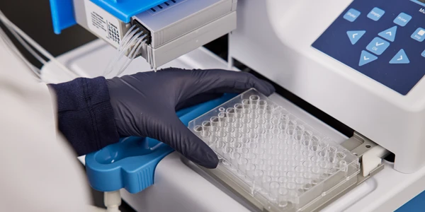
How Microplate Dispensers Enable Modern Research and Discovery
From drug discovery to genomics, almost every lab today depends on microplate dispensers.
Dec 08, 2025
Technical Insight
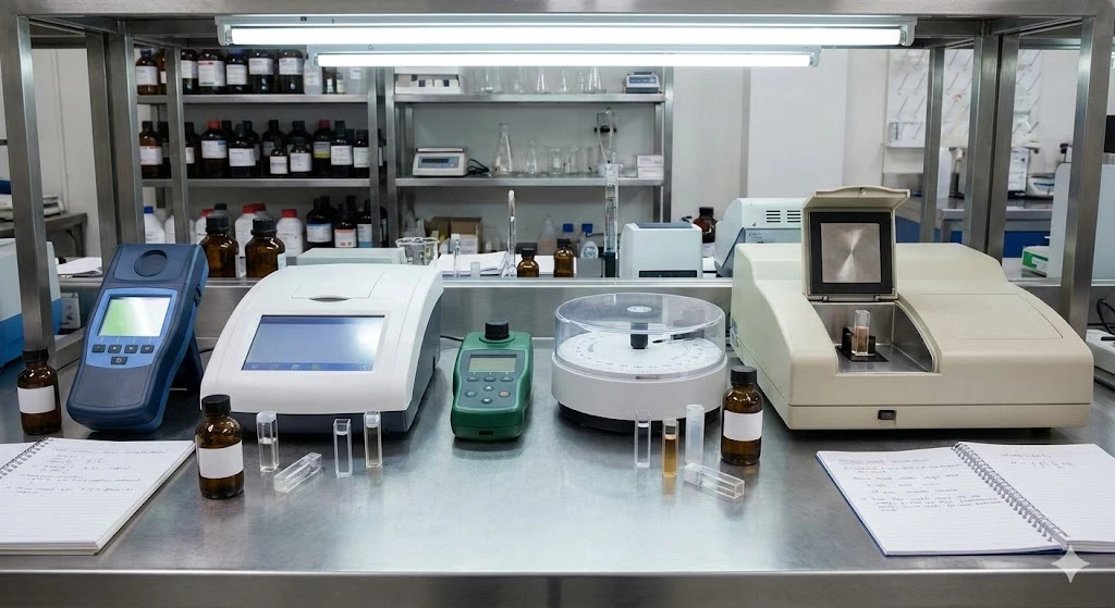
The Best Colorimeters of 2026
Discover the top-rated colorimeters of 2026 for laboratory professionals. We review the best new models for accuracy, speed, and chemical analysis.
Dec 08, 2025
Technical Insight
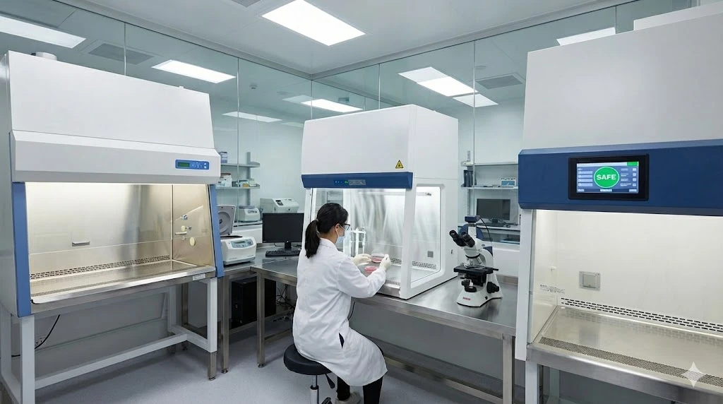
The Best Biosafety Cabinets and Laminar Flow Hoods of 2026
Discover the top biosafety cabinets and laminar flow hoods of 2026. This guide reviews safety, efficiency, and features for optimal laboratory protection.
Dec 05, 2025
Technical Insight
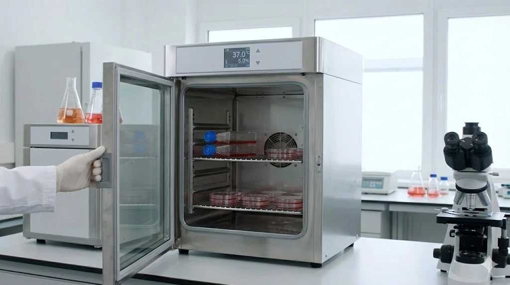
The Best Laboratory Incubators of 2026
Discover the best laboratory incubators of 2026. This guide reviews top models for cell culture and microbiology, focusing on precision, capacity, and new releases.
Dec 05, 2025
Technical Insight
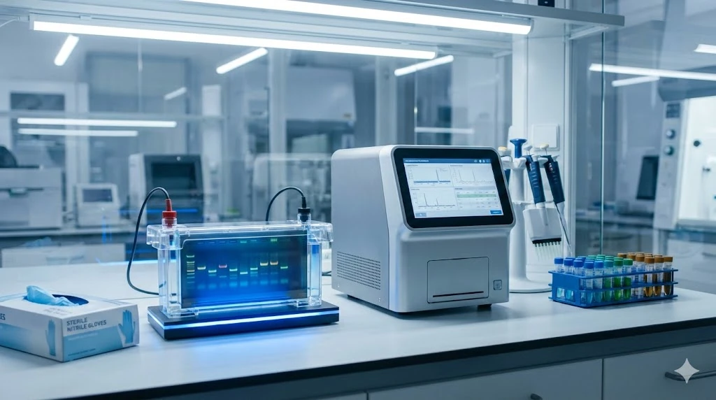
The Best Electrophoresis Equipment of 2026
Discover the top electrophoresis equipment of 2026 for research and clinical labs. Compare models for budget, throughput, and versatility.
Dec 05, 2025
Technical Insight
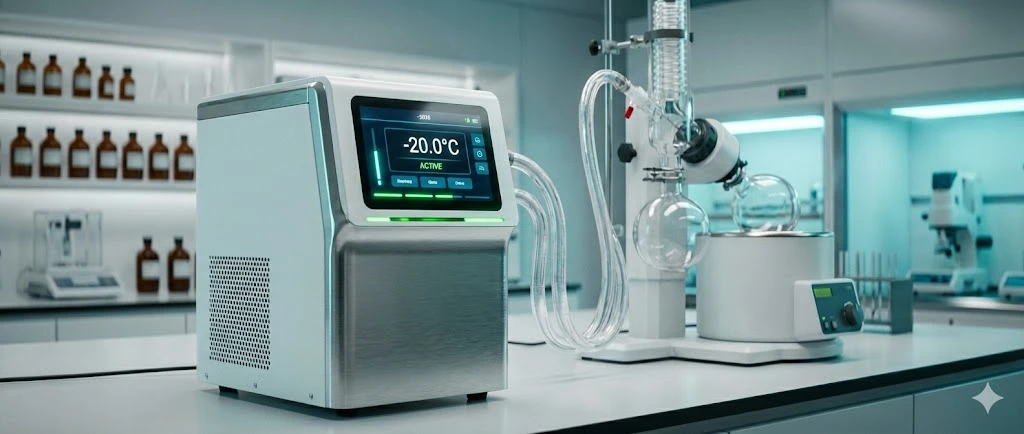
The Best Laboratory Chillers of 2026
Discover the top-rated cooling systems for 2026. This guide reviews high-performance, eco-friendly, and budget-conscious lab chiller models.
Dec 03, 2025
Featured, Popular Products
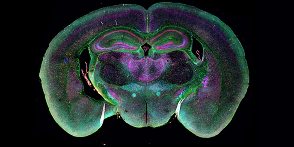
Remapping Research and Discovery with Spatial Imaging
By integrating molecular identity with spatial relationships, researchers can build more predictive models, uncover hidden cellular interactions, and generate insights that would remain invisible using conventional methods alone.
Dec 03, 2025
Featured, Popular Products
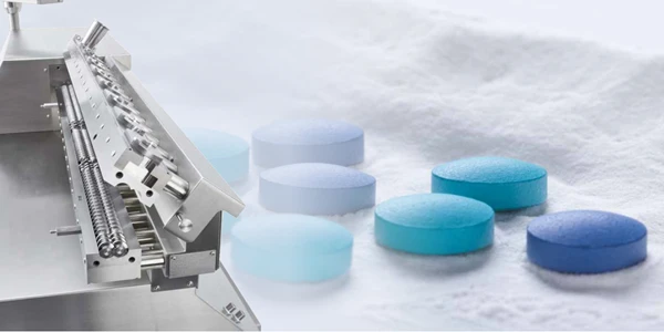
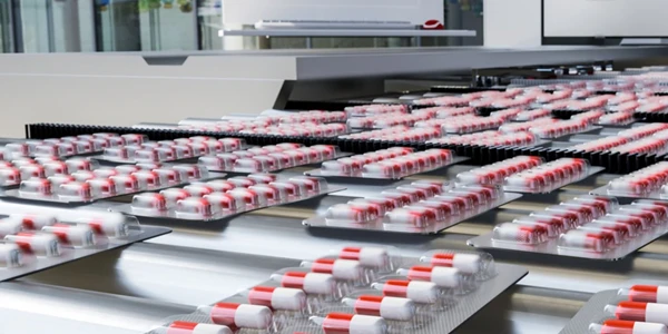
The Evolution of Continuous Processing in Pharmaceutical Development and Manufacturing
Ongoing advancements in technologies including hot-melt extrusion and twin-screw granulation will further accelerate the transition to continuous processing as the new standard in pharmaceutical development.
Dec 02, 2025
Featured, Popular Products
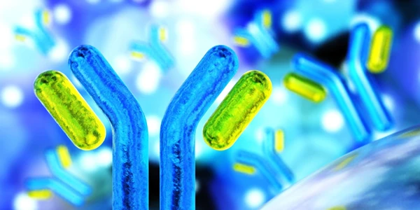
The Expanding Impact of Biolayer Interferometry on Antibody Development
Biolayer Interferometry (BLI) now sits at the center of modern antibody R&D - delivering kinetic, affinity, specificity, and competitive binding insights in a single streamlined platform.
Nov 24, 2025
Buying Guides, Featured
Advanced Raman Spectroscopy and the Need for Rapid and Reliable Data in Pharma Testing
As the pharma industry continues to shift toward agile manufacturing, continuous processing, and real-time release testing, advanced Raman spectroscopy technologies are set to lead the way.