ImageFX (2025)
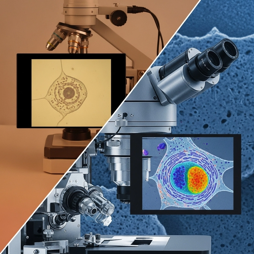
How does electron microscopy work?
Three main factors underlie the scientific principle of microscopy: magnification, illumination, and resolution.
- Resolution is perhaps the central tenet of microscopy in that it defines the ability of any device to distinguish between two closely spaced objects. In other words, the ability to reveal structural details as distinct and separate.
- Diffraction (or the mathematically defined Diffraction Limit) is used as the measure of resolution and is impacted by factors including: the wavelength of incident light, the refractive index of materials through which the light passes, and the numerical aperture of the lens used to magnify the image.
- Consequently, there is a limit (the Diffraction Limit) beyond which the ability to resolve two images becomes impossible. The lowest diffraction achievable with conventional lenses is 200 nm, and efforts to surpass this point have been limited even with modern techniques, although there are notable exceptions.
How do SEM electron microscopes work?
The electron microscope concept overcomes the diffraction limit and permits imaging of much smaller and more complex substances and biological specimens.
Scanning Electron Microscopy (SEM)
SEM involves the production of an image by scanning the specimen with a focused beam of electrons.
- These electrons interact with electrons in the sample and produce signals representative of the sample topography and composition.
- The beam position is closely linked with the emission signal to construct a resulting image in fine detail — at a resolution in the low nm range.
- The most commonly used method in biological applications is secondary electron detection from excited atoms within the beam.
- Others include back-scattered or reflected electrons and cathodoluminescence. It is rare, however, that a SEM instrument can detect more than one signal type simultaneously.
The image is produced from electronic interactions at various depths of the specimen, and secondary electron imaging (SEI), the most common method, is performed by detection at the very surface.
- The result is very high 1 nm surface resolution — although subsurface detail is precluded.
- To this end, back-scattered electrons (BSE), involving interactions deeper within the sample and through a separate electronic mechanism, can provide subsurface identification of the elemental composition of a sample.
What is a limitation of using SEM electron microscopes to view specimens?
The power of SEM does come with certain requirements for sample preparation.
- Specimens must be both electrically conductive at the surface and electrically grounded to prevent accumulation of static charge.
- Nonconductive samples can be coated with a conductive film such as gold or palladium.
- Alternatively, environmental or low-voltage can be used on native non-conducting samples, although these may require specific (and sometimes tedious) conditions.
- Biological samples have a requirement to be completely dry due to high vacuum conditions of the mounting chamber. Therefore, most samples involving live tissues and cells must be chemically fixed by treatment with glutaraldehyde or formaldehyde to preserve native structure.
- SEM samples can also be cold treated in a process called cryofixation (typically involving a SEM stage built for this) and can be imaged by low-temperature scanning.
- Samples can also be freeze-fractured to reveal inner structures in fine detail.
The resolution in SEM is not limited by the diffraction limit or the quality of lenses or mirrors, etc.
- Rather, spatial resolution in SEM depends on the size of the electron beam and the volume of specimen material interacting with the beam — or interaction volume.
- As a result, the resolution falls between 1 nm and 20 nm. Impressive indeed, although not as high resolution of the next technique to be discussed, which can measure distances between individual atoms in a sample.
How do TEM electron microscopes work?
Transmission Electron Microscopy (TEM)
TEM is similar to SEM in that a narrow beam of electrons is projected onto a sample.
- A major difference, however, is that the electronic interactions are captured after transmission through the thinly sliced sample, after which the signal is amplified and then interpreted to yield structural information.
- TEM is capable of imaging at the single electron level, many thousands of times smaller than light microscope limits.
- Various imaging modulations can be performed to account for the thickness and condition of the sample.
What is a limitation of using TEM electron microscopes to view specimens?
With regard to TEM:
- Preparation of cells for TEM requires fixation (with chemicals or cryotechniques) that can introduce artifacts and damage.
- The preparation of cells/tissues for TEM is lengthy.
- TEM relies on highly sophisticated and expensive machinery.
Regarding Cryo-TEM:
- The samples are unstained, therefore, the technique produces low-contrast images.
- Thin sectioning of samples in a frozen state is a technical and tedious procedure.
- Cryo-TEM relies on highly sophisticated and expensive machinery.
Two considerations dominate the discussion:
- Although a powerful, high resolution technique, traditional and cryo-TEM require extensive and tedious sample preparation. This often involves ultra-thin sectioning by use is a microtome and sample fixation, both of which can introduce artifacts and can damage specimens.
- The second consideration is the large investment in obtaining and maintaining a TEM instrument.
In the end, it is again time and money that seem to rule the discussion on limitations. The resolving power and unique capabilities of the technique, however, may very well negate these considerations.
Explore the full range of new and used electron microscopes at labx.com.