Advancements in Brain Imaging Technologies Showcased at the Society for Neuroscience Conference

Written By: Damon Anderson, PhD
Nov 27, 2018
Enhancing the range and scope of neural imaging
The annual meeting of the Society for Neuroscience is a humbling experience. The electrifying hum of high-minded neuro-speak echoes among the attendees – over 30,000 strong.
This year was exciting from numerous perspectives. A resounding topic of discussion this year included new imaging technologies and the insight these may provide into complex brain processes.
Integrated microscopy image analysis
Zeiss showcased several key developments and collaborations in the areas of multi-aspect imaging and data analysis. With a broad portfolio of instruments, Zeiss aims to empower investigations across all scales – from the submicron to the hundred-micron range and beyond. Critical to this vision are the levels of integration required in order to piece imaging techniques together to form larger, more comprehensive pictures of the brain and associated neural processes.
Zeiss ZEN Connect is an enhanced imaging software platform designed for advanced visual analysis. Built to ensure users focus on the data rather than the acquisition method, ZEN Connect enables workflows initiated on any microscope, throughout multiple ranges of magnification, to be automatically overlaid and relocated using mapping algorithms. The technology supports data analysis in a wider context, connecting large field of view images with high resolution details.
- ZEN Connect is available for the company’s full range of optical, confocal, X-ray, electron, and ion microscopes.
- The software is offered on a limited trial basis to allow users to sample the product – available online from the Zeiss microscopy webpage.
- Existing customers can use it in conjunction with the ZEN Blue or SmartSEM 6.2 or higher software, while new users can download ZEN Lite to test the capabilities of ZEN Connect.
Brain imaging in live animals in real time
Also announced at the Zeiss press conference was an interesting collaboration with Inscopix, a company focused on mesoscale imaging and brain mapping technologies. Inscopix has pioneered miniature head-mountable microscope technology which allows imaging of large-scale brain circuit dynamics -- in live animals and in real time. These “portable” microscopes are connected to controllers which can modulate activities such as calcium signalling in discrete areas of the brain during natural behavior.
Coupled with the Zeiss LSM800 and Airyscan platforms, the system targets image resolution enhancement of brain circuit activity for research and drug development.
3D analysis of complex structures
Olympus showcased a range of microscope platforms and imaging software solutions, including several recent product releases.
NoviSight cell analysis software is designed to provide statistical data for complex samples, spheroids, and other 3D objects in microplate-based experiments. The technology allows users to check the morphology of samples by measuring a range of spheroid or cell nuclei parameters. The software makes it possible to measure and analyze physiological 3D models, helping improve the speed of HCS processes and drug discovery.
- Images are captured in OIR and VSI formats
- Compatible with microplate: 6, 12, 24, 48, 96, 384 wells
- Image views: 2D view (Single/Three Slides/MIP), 3D view (Isosurface/MIP/Alpha Blend)
- Graph Views: Histogram or Scattergram
- Analysis/Statistic: Various morphological measurement, Table view, Gating, Gallery
- Options: Recognition option, Measurement option, Statistical option
Labeling and high-speed imaging of intact tissue
Combining the themes of enhanced image resolution with 3D sample analysis, Life Canvas Technologies brings an innovative perspective to whole organ visualization through their suite of imaging solutions. The company’s technology portfolio includes three platforms designed to enhance and enable visualization of 3D structures and molecular phenotyping in the brain.
- Clarity is a technique that enables the transformation of intact tissue into a nanoporous hydrogel-hybridized form (cross-linked to a 3D network of hydrophilic polymers), that is fully assembled but optically transparent and semipermeable.
- SWITCH introduces a simple method that enables proteomic imaging for scalable, integrated, high-dimensional phenotyping of both animal tissues and human clinical samples.
- Stochastic Electrotransport introduces a novel electrostatic method for rapid non-destructive processing of porous samples.
This latter method forms the basis of the SmartLabel instrument platform, which enables complete clearing of model organs within 1-3 days and staining with nuclear dyes, proteins, and antibodies in a 1-day period.
- The SmartLabel device performs an order of magnitude faster than passive labeling – 1-3 days versus weeks or months.
- The device is easy to use and reliable – preventing tissue damage via electrotransport technology, with minimal probe loss and tissue contamination using nanoporous membranes.
- The platform offers a complete and uniform staining solution with low consumption of antibody compared with alternative techniques.
The SmartSPIM instrument platform is an advanced light sheet microscope capable of imaging large tissue samples with high speed and resolution. The unique illumination arm of the instrument creates a flat-top illumination profile laterally, and a focused beam axially. The illumination optics scan the beam axially while synchronized with the imaging camera.
- SmartSPIM is capable of imaging a wide range of sample sizes
- The instrument provides high axial resolution with many different objectives
- Uniform axial resolution is provided across entire samples
Perhaps the most interesting aspect is the high-speed volumetric imaging capabilities, allowing image capture of a mouse brain hemisphere in less than 15 minutes or a whole mouse brain in less than 25 minutes at 1.53 µm x 1.75 µm x 4 µm voxel size.
Progress in brain mapping and visualization
These are a mere few of the advancements offering insight into brain imaging, brain mapping, and neural circuitry visualization. The broad spectrum of powerful new technologies on display this year offers promise towards a greater understanding of the world’s most complex biological system.
Jun 27, 2025
Featured, Product News, Technical Insight
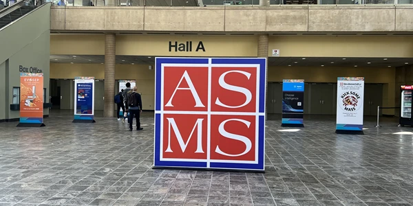
2025 ASMS: New Mass Spectrometry Technologies to Confront the Next Wave of Complex Challenges
The year’s ASMS conference showcased new technologies that expand analytical capabilities while refining size and workflow efficiency

Jun 25, 2025
Product News
Bio-Rad Expands Bioanalytical Antibodies Portfolio with New Anti-Idiotypic Tools and SpyCatcher Reagent
Bio-Rad introduces new bioanalytical antibodies and IgM-FcSpyCatcher, expanding options for drug monitoring and assay flexibility in research labs.

Jun 24, 2025
Product News
Keyence Launches BZ‑X1000: High‑Res, All‑in‑One Fluorescence Microscope
Keyence’s BZ‑X1000 delivers 3.5× higher resolution, 5× faster imaging, and modular, multi-modal fluorescence capabilities—all on a bench-top system.

Jun 23, 2025
Product News
From Delivery to Disposition
reLink Medical adds medical equipment procurement, rental, and flat-rate repair options to industry leading platform
Jun 20, 2025
Product News
Enhanced Vanquish CAD P Series Advances Impurity Analysis Performance
Explore the Vanquish CAD P Series for high-sensitivity impurity analysis with better reproducibility and detection of semi-volatile compounds.
Jun 20, 2025
Featured, Popular Products
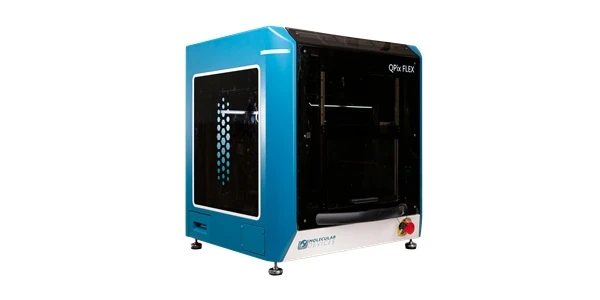
Navigating the Challenges of Microbial Research
Learn how the QPix® FLEX™ system transforms microbial research by automating colony picking, plating, liquid handling, and streaking – all on a single instrument.
Jun 18, 2025
Product News
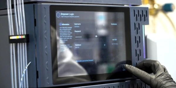
Alliance iS HPLC Software 2.0 Sets New Benchmark for HPLC Software Security
Waters Alliance iS HPLC Software 2.0 boosts traceability and touchscreen security in HPLC software, helping labs meet FDA data integrity standards.
Jun 16, 2025
Product News
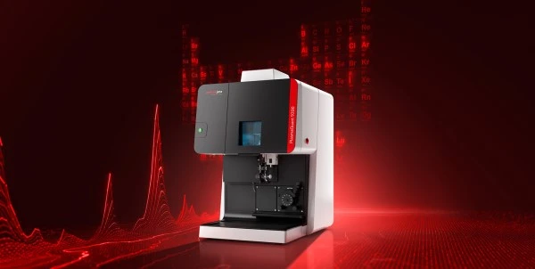
Discover the PlasmaQuant 9200: High-Resolution ICP-OES
Explore Analytik Jena's PlasmaQuant 9200 ICP-OES—high-resolution elemental analysis with compact design and superior matrix tolerance.
Jun 10, 2025
Product News
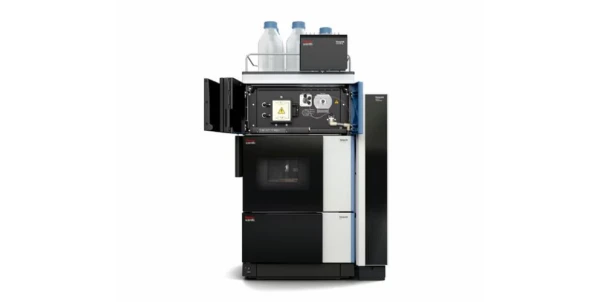
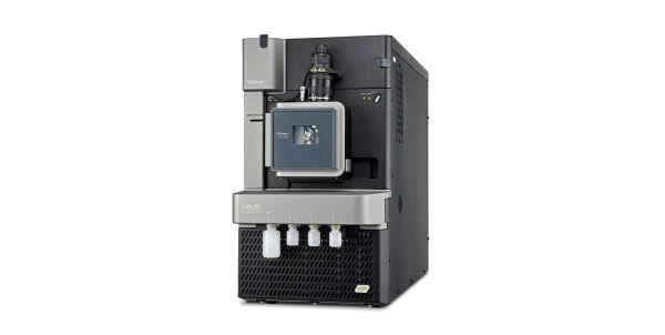
Waters Xevo TQ Absolute XR Mass Spectrometer Sets New Benchmark
Discover the Xevo TQ Absolute XR Mass Spectrometer—Waters' most advanced benchtop tandem quadrupole for PFAS detection and trace-level analysis.
Jun 06, 2025
Product News
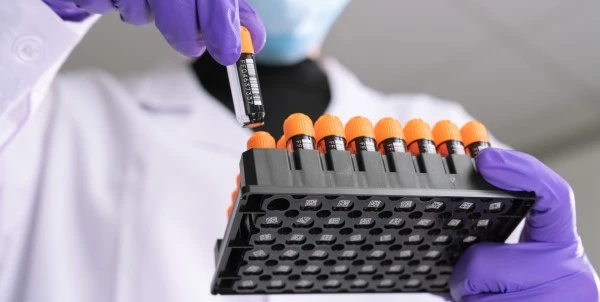
Azenta Introduces E-Beam Sterilized FluidX Tubes
Azenta's new E-Beam treated FluidX tubes offer chemical-free sterilization for secure sample storage in various laboratory applications.