New Technologies Enhance the Speed and Accuracy of Flow Cytometry Workflows

Written By: Damon Anderson, PhD
Mar 06, 2019
Flow cytometry has become an essential tool for separation and characterization of single cells in heterogeneous populations. Immune cell and stem cell classification for research and clinical applications are examples, among a wide array of applications. View a brief primer on Flow Cytometry.
Drawbacks of the technique traditionally included relatively low-throughput workflows, arising from limited multiplexing and sample processing speeds. Technical limitations affecting accuracy, as well as complex operation, have also been difficulties in the past.
State-of-the-art flow cytometry systems are harnessing multiplexed detection and automatable workflows, significantly enhancing the scale and speed of analysis.
Advanced focusing technologies are boosting the strength and accuracy of cellular characterization – with overall implications for the fields of immunology, oncology, cell therapy, and other areas of clinical importance.
Flow cytometry protocol
At the basic level, flow cytometry can detect the volume, cell-size, and structural complexity of cells, typically by capturing side and forward scattering light, or fluorescence, projected by a laser light source on a flow cell. The presence of specific DNA, RNA, protein, antibodies, etc. requires use of fluorophore conjugated antibodies and lasers of specified wavelengths.
The sample flow cell is typically attached to sensitive detectors to capture the intensity of emitted light on fast time scales. These signals are then used to deduce the state of the cells or internal components relative to blanks or control cells.
Traditional workflows are complex, sometimes prone to variability in sample prep -- and some markers are cytotoxic, further challenging accuracy. Overall, the analysis time can be lengthy, especially with large cell populations and mixed phenotypes.
New flow cytometry technologies
Three main aspects of technology development are together having significant impact of the power and performance of modern flow cytometry instrumentation.
Multiplex flow cytometry
The advent of multicolor flow cytometry has led to an evolution in the workflows of the traditional technique.
The number of targets which can now be measured in a given sample or experiment has increased and is now limited only by the number of light sources and detectors available on a given instrument.
The use of multiple laser light sources can be coupled with fluorophores with distinct absorption profiles – expanding the number of antibodies and targets available for measurement.
Multiple lasers can also be used in separate channels or in tandem to measure differing absorption profiles within the same sample or cell.
The use of multiple detectors enables the measurement of emitted light covering a broad spectrum of fluorophore signals – again individually, in combination, or in sequence.
The net benefit of these advancements is the ability to now measure, for instance, cellular effects of experimental stimuli, the origin or fate of cellular features over time, or the coexistence of multiple immune markers.
Multiplexed detection conitnues to evolve with a large array of adaptable configurations now available.
Flow cytometry processing speed and automation
High-throughput processing is another technology feature driving the evolution of flow cytometry instrumentation.
Traditional instruments have been limited by, not only the number and arrangement or light sources, detectors, and flow cells, but by the inability to interface with automation.
Even high-end, multifunctional instruments, while powerful, have traditionally been restricted by the operator and sample interface, as well as data analysis and management at the tailend.
Upgrades to the sample interface and fluidics handling systems has now enabled more streamlined coupling of automation modules.
Plate robots, plate hotels, incubators, and reagent handling products can together serve to significantly increase the rate of sample preparation.
The ability to process 96- or 384-well plates (or more) has clear implications on processing rates for sample intensive applications.
Control software to manage all of these features, with the ability to interface directly with laboratory information management systems (LIMSs), creates a central command interface – decreasing complexity of operation and increasing workflow efficiency.
In total, these advanced features should now support large-scale, high-throughput applications such as biomarker discovery and phenotypic screening – workflows which were unachievable with previous flow cytometer technologies.
Advanced flow cytometry focusing and accuracy
A third enhancement for modern flow cytometer performance involves increased accuracy arising from new focusing technologies.
Acoustic focusing involves the use of ultrasound waves to funnel cells into and through the flow cell and detection point. The advantages of this approach over the more typical liquid flow cell alignment includes higher throughput processing of more dilute samples or populations of specific cells, such as stem cells, in heterogenous samples.
Although in the experimental stage, combined ultrasound (US) backscatter and photoacoustic (PA) (combined acoustic and laser irradiated) approaches will theoretically enable single cell in vitro label-free multiparametric analysis based on two difference physical interactions. Photoacoustic signals contain information about the cellular components such as lipids, mitochrondria, DNA, etc. Ultrasound backscatter collects information on cell shape, size, and biomechanical properties.
Together, these powerful techniques may promise to significantly increase the accuracy and scope of flow cytometry applications.
Summary
Technology development is moving quickly to keep pace with modern research and clinical approaches towards immunotherapy and cell therapy in a variety of disease scenarios.
It will be fascinating watch as these new technologies continue to expand and flow into the marketplace.
Feb 12, 2026
Product News
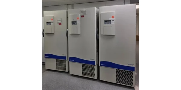
$5M+ in GMP Biopharma Manufacturing & QC Equipment Heads to Online Auction
A Southern California GMP manufacturing and clinical-stage QC laboratory is liquidating new and late-model equipment and unused inventory in an online auction opening February 11, 2026

Feb 12, 2026
Product News
2026 SLAS: Inspiration Catalyzing Discovery
A LabX look at the annual conference and exhibition, a key forum for networking, collaboration, and advancement of new technologies
Feb 11, 2026
Product News
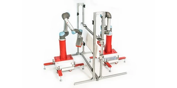
Angstrom Scientific Named U.S. Distributor for Radalytica’s RadalyX Robotic X-ray and CT System
Angstrom Scientific, known for representing microscopy and materials analysis technologies across North America, adds Radalytica’s robotic X-ray and CT platform to its portfolio of analytical and imaging solutions.
Feb 10, 2026
Technical Insight
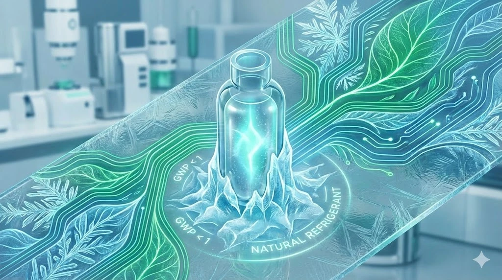
Eco-Friendly Refrigeration: Evaluating Natural Refrigerants and Low-GWP Cooling Systems
This guide explains the operational and environmental benefits of adopting natural refrigerants and low-GWP cooling systems in laboratory settings
Feb 10, 2026
Technical Insight
The Ultimate Guide to Sustainability in Laboratory Equipment and Operations
A comprehensive analysis of sustainable laboratory practices, focusing on energy-efficient equipment, waste management, and digital workflows

Jan 09, 2026
Product News
BRANDTECH Scientific Introduces the Transferpette® pro Micropipette: A New Twist on Comfort and Control
The latest addition to its trusted line of precision liquid handling instruments, engineered for comfort, control, and repeatable accuracy.

Jan 06, 2026
Featured, Technical Insight
New Research Frontiers Unlocked by Next-Generation Flow Cytometry
Next-generation flow cytometry systems using acoustic focusing are opening entirely new areas of research that were previously impractical or inaccessible with conventional flow cytometers.

Jan 05, 2026
Buying Guides
The Bottom Line: How Smart Microcentrifuge Tube Choices Impact Lab Efficiency and Cost
Innovative microcentrifuge tube solutions are more than just lab consumables—they’re strategic tools that can reduce costs, improve outcomes, and support long-term sustainability goals.

Jan 05, 2026
Buying Guides
Future-Proofing Your Lab with Innovative Microcentrifuge Tube Solutions
As laboratories evolve to meet the growing demands of modern science, even the most basic tools—like microcentrifuge tubes—are being reimagined.

Jan 05, 2026
Buying Guides
Microcentrifuge Tube Innovations: Driving Sustainable Change
Integrating next-generation consumables into standard workflows can lead to a more sustainable and responsible research environment.