Single-Channel vs. Multi-Channel Electrophysiology: A Comprehensive Guide

Written By: Craig Bradley
Nov 06, 2019
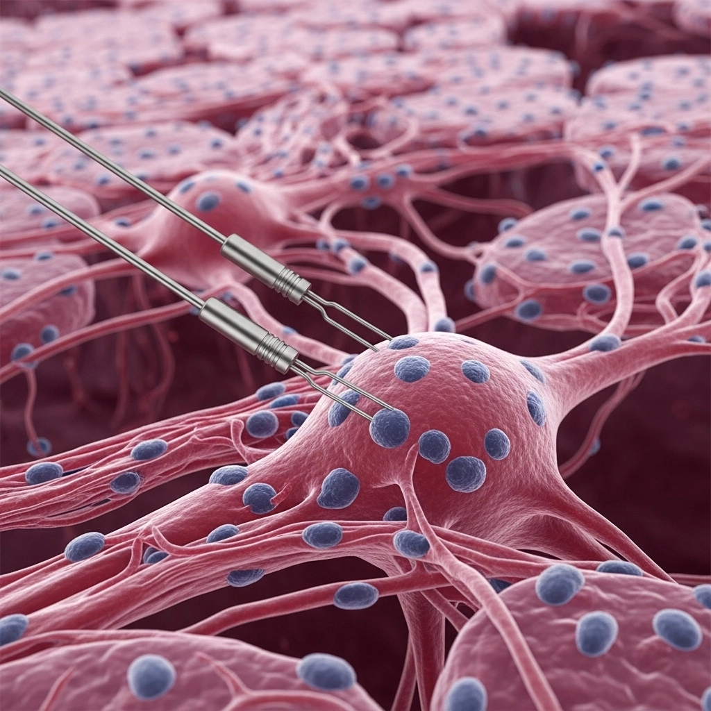
ImageFX (2025) In the intricate world of neuroscience and cellular biology, understanding the electrical language of cells is paramount. Electrophysiology, the study of these bioelectrical signals, provides invaluable insights into cellular function, disease mechanisms, and drug interactions. Whether you're a seasoned electrophysiologist or new to the field, navigating the nuances of single-channel versus multi-channel recording techniques can be complex. This guide aims to demystify these powerful methods, offering a clear comparison to help you choose the optimal approach for your research. At its core, electrophysiology investigates the voltage differences, or potentials, that exist across the membranes of excitable cells. These include vital cell types such as cardiac myocytes, sensory neurons, and motor neurons, all of which exhibit dynamic electrical activity. The movement of ions across the cell membrane, facilitated by specialized voltage-sensitive channels, generates measurable electrical currents. By studying these currents, researchers can decipher how cells respond to various stimuli, from neurotransmitters to pharmaceutical compounds. Key techniques used to measure these electrical phenomena include: Voltage Clamp: This technique allows researchers to maintain a cell's membrane potential at a constant, chosen value. By doing so, it becomes possible to precisely measure the ion currents flowing across the membrane in response to channel activation or de-activation. This is crucial for understanding the kinetics of ion channel function and how drugs or mutations might alter them. Current Clamp: Conversely, current clamp involves injecting a specific current into the cell and measuring the resulting changes in membrane voltage. This method is vital for observing how voltage-gated channels activate in response to ion flux, action potentials, and the influence of neurotransmitters, providing insights into a cell's natural electrical behavior. Various recording setups facilitate these measurements, each with unique applications: Intracellular Recording: A microelectrode is carefully inserted into the cell to measure voltage and/or current directly from the intracellular compartment, compared against a reference electrode in the bathing solution. Patch Clamp Recording: This advanced technique involves forming a tight seal between a glass electrode and a small "patch" of the cell membrane. Gentle suction allows for precise measurements of ion flow across this isolated membrane section. Whole-Cell Recording: An extension of patch clamp where increased suction ruptures the membrane patch, providing access to the entire cell's interior and allowing measurement of total cellular voltage or conductance. Excised Patch Recording: Here, the membrane patch is completely detached from the cell while remaining sealed to the electrode. This allows for manipulation of both the intracellular and extracellular sides of the membrane, offering high sensitivity for studying channel activity. Artificial Lipid Bilayers: Channels can be incorporated into a synthetic lipid membrane formed across a tiny aperture, allowing for controlled study of individual channel behavior in a simplified environment. Multi-channel electrophysiological recordings represent the collective measurement of electrical activity from a population of ion channels across a cell membrane. Imagine it like listening to a piano chord – you hear a rich, collective sound produced by multiple notes playing simultaneously. This approach is instrumental for understanding the overall electrical behavior of a cell in response to various stimuli. Applications and Advantages: Holistic Cellular Response: Multi-channel recordings are invaluable for studying how an entire cell reacts to chemical or electrical stimuli, such as drugs, neurotransmitters, or changes in mechanical force. Drug Discovery and Toxicology: These recordings are fundamental in assessing the impact of compounds on cellular activity, crucial for pharmaceutical development and understanding toxicological effects. Kinetic Analysis: Researchers can meticulously examine the kinetics of channel opening, closing, de-activation, and blockage, providing detailed time- and concentration-dependent insights. Cell Models: To study specific channel activity, target proteins are often overexpressed in suitable cell backgrounds. Xenopus oocytes, large unfertilized frog germ cells, are a popular system due to their ease of manipulation for DNA/RNA injection and electrode insertion. Limitations: Population Average: The primary limitation is that measurements reflect the average activity of many channels. This means individual channel attributes, such as the precise conductance of a single open channel, flickering events, or subtle blockages, cannot be directly observed. Masked Heterogeneity: It can be challenging to discern if observed conductance changes are due to the physical dimensions of the channel or rapid opening/closing events. Furthermore, multi-channel recordings may obscure heterogeneity within a channel population, where some channels might function aberrantly while others perform normally. In contrast to the collective view of multi-channel recordings, single-channel electrophysiology provides discrete, high-resolution measurements of individual ion channel activity. This is akin to isolating a single note from a piano chord – allowing you to analyze its unique frequency and amplitude. This technique offers unparalleled detail into the fundamental behavior of ion channels. Advantages: High-Resolution Insights: Enables the observation of discrete "jumps" in conductance as a single channel opens and closes, providing direct evidence of individual gating events. Detailed Gating Kinetics: Researchers can precisely measure the duration of individual channel opening and closing events, as well as the frequency of channel gating. Direct Observation of Blockage: Allows for the direct visualization of channel blockage by exogenous compounds or de-activation due to overstimulation, providing clear mechanistic insights. Understanding Channel Flickering: Unique events like channel flickering, which are masked in multi-channel recordings, become clearly observable. Challenges and Limitations: Channel Isolation: A significant challenge lies in balancing channel expression levels with the ability to isolate individual channels within a patch. Too high expression makes isolation difficult, while too low makes channels hard to locate. Recording Environment Sensitivity: Single-channel recording demands an exceptionally controlled environment, free from vibrational or electrical noise, which can easily obscure subtle single-channel events. Membrane Integrity: The integrity of the cell membrane or artificial bilayer is critical; any compromise or contamination can severely impact observations. Artificial Bilayer Considerations: While artificial bilayers offer excellent control over channel insertion, the absence of native cofactors or modulating proteins must be considered, as this can affect channel activity compared to a physiological context. Both single-channel and multi-channel electrophysiology offer unique strengths and provide complementary insights into cellular processes. Multi-channel recordings excel at capturing the overall physiological response of a cell, making them ideal for drug screening and understanding population-level effects. Single-channel recordings, on the other hand, provide the granular detail necessary to unravel the fundamental biophysics of individual ion channels. Often, combining both approaches yields the most comprehensive understanding, allowing researchers to move from the macroscopic cellular response to the microscopic behavior of individual molecules. The choice of technique ultimately depends on your specific research question and the level of detail required. Regardless of your focus, having access to reliable electrophysiology equipment and resources is crucial for successful experimentation. Explore the cutting-edge electrophysiology solutions and resources available at LabX to advance your neuroscience research. What is the primary difference between single-channel and multi-channel electrophysiology recordings?
Single-channel recordings measure the discrete electrical activity of individual ion channels, showing distinct opening and closing events. Multi-channel recordings, conversely, capture the summed electrical activity from a population of many ion channels, providing an overall cellular response. Why are Xenopus oocytes commonly used in electrophysiology research?
Xenopus oocytes are favored as a cell model because they are large, unfertilized germ cells that are easy to manipulate. They can be readily injected with DNA or RNA to overexpress specific ion channels, making them ideal for studying the function of these channels in a controlled environment. What are the main advantages of using patch clamp techniques in electrophysiology?
Patch clamp techniques offer high-resolution measurement capabilities, allowing researchers to study ion currents across very small patches of membrane or even individual channels. This method provides precise control over the membrane potential and allows for detailed kinetic analysis of channel behavior. How do voltage clamp and current clamp techniques differ in electrophysiology?
Voltage clamp techniques hold the membrane potential constant to measure ion currents, useful for studying channel kinetics. Current clamp techniques, however, inject current into the cell and measure changes in membrane voltage, which is essential for understanding how channels contribute to action potentials and overall cellular excitability.
Understanding Electrophysiology: The Basics of Cellular Electrical Activity
Multi-Channel Electrophysiology: Capturing the Symphony of Cellular Signals
Single-Channel Electrophysiology: Unveiling Individual Ion Channel Dynamics
Choosing the Right Electrophysiology Technique for Your Research
Frequently Asked Questions
Mar 02, 2026
Product News
Thermo Fisher Launches Fluid Ease™ Pro ClipTip™ Electronic Pipette with High-Resolution Touchscreen and Advanced Workflow Controls
The system combines programmable control, intelligent power management, and a secure tip attachment mechanism to support consistent, reproducible liquid handling.
Feb 26, 2026
Product News
Waters Launches Microflow LC Columns with MaxPeak Premier Technology for Higher Sensitivity and Lower Resource Consumption
The product launch reflects continued investment in chromatographic chemistries that address real-world bottlenecks in method development and routine analysis
Feb 19, 2026
Technical Insight
Implementing Hydrogen Generators for Clean Lab Gas Supply: Safety and Benefits
Transitioning to on-site gas production streamlines analytical workflows, eliminates hazardous cylinder storage, and supports facility sustainability.
Feb 19, 2026
Technical Insight
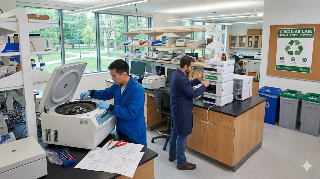
Circular Economy in Laboratory Equipment: How to Adopt Repair, Reuse, and Refurbishment Strategies
Adopting a circular economy for laboratory equipment minimizes environmental impact while maximizing instrument lifespan and optimizing laboratory budgets.
Feb 12, 2026
Product News
$5M+ in GMP Biopharma Manufacturing & QC Equipment Heads to Online Auction
A Southern California GMP manufacturing and clinical-stage QC laboratory is liquidating new and late-model equipment and unused inventory in an online auction opening February 11, 2026

Feb 12, 2026
Product News
2026 SLAS: Inspiration Catalyzing Discovery
A LabX look at the annual conference and exhibition, a key forum for networking, collaboration, and advancement of new technologies
Feb 11, 2026
Product News
Angstrom Scientific Named U.S. Distributor for Radalytica’s RadalyX Robotic X-ray and CT System
Angstrom Scientific, known for representing microscopy and materials analysis technologies across North America, adds Radalytica’s robotic X-ray and CT platform to its portfolio of analytical and imaging solutions.

Feb 10, 2026
Technical Insight
Eco-Friendly Refrigeration: Evaluating Natural Refrigerants and Low-GWP Cooling Systems
This guide explains the operational and environmental benefits of adopting natural refrigerants and low-GWP cooling systems in laboratory settings
Feb 10, 2026
Technical Insight
The Ultimate Guide to Sustainability in Laboratory Equipment and Operations
A comprehensive analysis of sustainable laboratory practices, focusing on energy-efficient equipment, waste management, and digital workflows
Jan 12, 2026
Technical Insight
The Best Polarimeters of 2026
A comprehensive guide to the top-rated polarimeters of 2026, featuring high-speed, high-precision, and budget-friendly models for every laboratory application.