Advances in Digital Imaging for Multiplex Western Blotting

Written By: Craig Bradley
Sep 26, 2025
GEMINI (2025)
The transition from single-target detection to simultaneous, multi-protein analysis is revolutionizing molecular biology, making multiplex western blotting an indispensable technique for studying complex cellular pathways. Traditional methods, often reliant on endpoint measurements, struggled to provide the quantitative accuracy and throughput required by modern research. The advent of sophisticated digital imaging systems has resolved these limitations, offering a wide linear dynamic range and the capacity to analyze multiple proteins, or targets, within a single blot. Achieving high-quality, quantitative data necessitates a deep understanding of the imaging hardware, reagent selection, and standardized analysis protocols. This article explores the core technological shifts and best practices driving precision in advanced western blotting workflows.
The Transition to Fluorescence for Multiplex Western Blotting
Historically, western blotting relied heavily on chemiluminescence (CL) for detection. While CL offers high sensitivity, its inherent limitations severely restrict quantitative multiplex western blotting. CL signals are transient, decaying rapidly, which makes accurate time-course comparisons and subsequent quantification challenging. Moreover, the signal saturation associated with CL often results in a narrow linear dynamic range (LDR), preventing accurate measurement of protein targets expressed across a broad concentration spectrum. This often forces the user to rely on inefficient and time-consuming stripping and reprobing protocols to assay multiple targets.
The adoption of fluorescence has transformed the landscape of digital imaging for western blotting. Fluorescence-based detection utilizes secondary antibodies conjugated to spectrally distinct fluorophores, such as those emitting in the near-infrared (NIR) region (typically
Stable Signal: Fluorescent signals are stable over time, allowing for image capture and re-analysis days after the primary experiment, ensuring reproducible data capture.
Wide Linear Dynamic Range: Digital imaging systems coupled with fluorescence allow for accurate protein quantification across a far wider range of concentrations compared to CL, minimizing saturation issues.
True Multiplexing: By using two or more fluorophores with non-overlapping emission spectra, two or more targets (e.g., a protein of interest and a loading control) can be assayed simultaneously on the same blot. The ability to perform simultaneous detection ensures that the signals from different targets are normalized to the same biological sample.
This shift enables higher throughput, reduces sample consumption, and inherently improves data quality by eliminating the variability introduced by stripping and reprobing steps.
High-Resolution Digital Imaging System Requirements
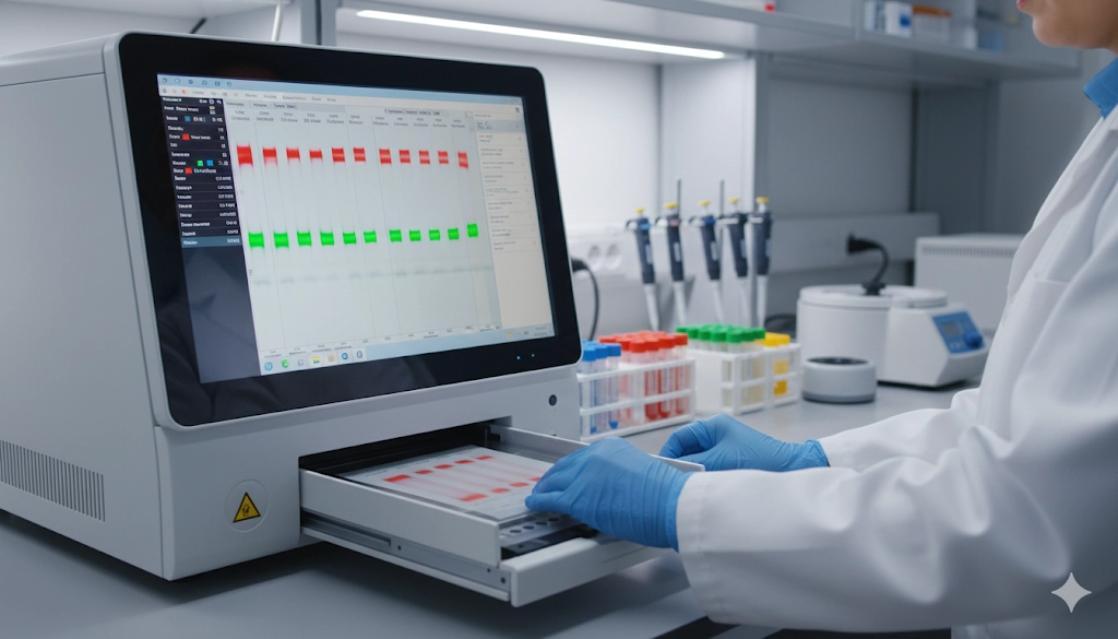
The successful execution of multiplex western blotting is reliant on the capabilities of the digital imaging system, which must be engineered to differentiate and capture distinct fluorescent signals with exceptional sensitivity and minimal noise. The critical components of these systems work in concert to achieve high-resolution nucleic acid imaging and protein quantification.
Central to the system is the combination of excitation sources and optical filters. Excitation is typically provided by highly controlled lasers or light-emitting diodes (LEDs) tuned to the specific excitation wavelength of the fluorophores in use. For example, a
The camera, often a cooled scientific-grade CCD or sCMOS sensor, is critical for capturing the resultant emission. These cameras feature high Quantum Efficiency (QE), meaning they are highly efficient at converting incoming photons into measurable electrical signals, which is vital for detecting low-abundance proteins. Crucially, the camera system must incorporate precise emission filters. These filters are engineered to allow only the specific fluorescence wavelength (e.g.,
Component | Function in Digital Imaging | Quantitative Benefit |
|---|---|---|
Laser/LED Excitation | Provides stable, monochromatic light for fluorophore activation. | Ensures consistent signal generation across the blot. |
Cooled Scientific Camera | Captures faint fluorescent light with high sensitivity and low thermal noise. | Extends the Linear Dynamic Range (LDR) for accuracy. |
Emission Filters | Isolates specific fluorescence signals (channels) based on wavelength. | Eliminates signal crosstalk between different target proteins. |
The superior resolution and low background noise offered by dedicated digital imaging systems are indispensable for accurate, quantitative multiplex western blotting.
Principles of Quantitative Multiplex Analysis
The primary advantage of using digital imaging for multiplex western blotting is the transition from qualitative band presence to true quantitative western blotting. This shift requires rigorous adherence to principles of signal linearity, saturation control, and robust normalization.
Achieving accurate quantification relies on capturing the signal within the assay’s Linear Dynamic Range (LDR). The LDR is the range of protein concentration where the detected signal intensity is directly proportional to the amount of protein present. Signals outside this range (i.e., saturated or too faint) cannot be used for accurate quantification. Modern digital imaging software often includes tools that automatically measure the pixel intensity across the entire blot and flag areas that are approaching or reaching saturation, guiding the user to adjust exposure times before data acquisition.
Normalization is arguably the most critical step in quantitative analysis. Small variations in sample loading, protein transfer efficiency, and well-to-well differences on the gel are inevitable. These variations must be corrected to determine the true relative change in protein expression.
Traditional normalization relied on housekeeping proteins (HKPs) such as
TPN uses fluorescent stains (e.g., those activated by specific NIR wavelengths) that bind to all proteins on the membrane. This total protein signal provides an accurate baseline against which the target protein signal is normalized. The calculation is performed simultaneously within the multiplex western blotting protocol:
This normalization strategy corrects for all pre-detection variability, ensuring that changes in the normalized target signal truly reflect biological changes in the sample.
Workflow Optimization for Reproducibility in Digital Imaging
To fully leverage the quantitative power of digital imaging in multiplex western blotting, every step of the laboratory protocol must be standardized. Discrepancies in blotting technique, membrane choice, or antibody titrations introduce confounding variability that no imaging system can entirely correct.
Key areas for workflow standardization include:
Antibody Titration: Both primary and fluorescently labeled secondary antibodies must be empirically titrated to ensure optimal signal-to-noise ratio. Over-titration leads to high background noise and faster signal saturation, compromising the LDR.
Blocking Buffer Selection: The choice of blocking agent (e.g., milk, bovine serum albumin, or proprietary formulations) is critical for minimizing non-specific binding of antibodies, which manifests as background fluorescence. The blocking buffer must be compatible with the fluorescent dye and the primary antibody.
Transfer Efficiency Assessment: While TPN corrects for loading variation, maximizing transfer efficiency remains important. Fluorescent TPN stains can be used immediately post-transfer to visually verify uniform protein migration and transfer quality before proceeding with immunodetection.
System Maintenance: Regular calibration of the digital imaging system's light sources and camera focus is required to maintain peak performance. Consistent cleaning of the sample stage and filter windows prevents light scatter and imaging artifacts.
A dedicated Standard Operating Procedure (SOP) that includes precise reagent concentrations, incubation times, and system settings is essential for achieving the inter-experiment and inter-user reproducibility required for high-impact research.
Maximizing Quantitative Accuracy in Western Blotting
The integration of specialized fluorescent reagents and high-resolution digital imaging systems has transformed western blotting from a semi-quantitative technique into a robust, high-fidelity quantitative assay. By adopting fluorescence over chemiluminescence, implementing rigorous normalization protocols, and adhering to strict workflow standardization, laboratories can maximize the quantitative accuracy of their protein analysis. This systematic approach to multiplex western blotting is vital for generating the verifiable and reproducible data required to advance biomedical research and diagnostics.
FAQ: Common Digital Imaging and Multiplex Western Blotting Questions
What is the significance of the linear dynamic range (LDR) in quantitative western blotting?
The LDR is the range where the detected fluorescent signal is directly proportional to the amount of protein, making it essential for accurate and reliable quantification of target protein expression.
How many targets can typically be analyzed in a single multiplex western blotting experiment?
Most standard digital imaging systems are optimized for two-color detection (two distinct near-infrared channels), allowing the simultaneous analysis of two targets, typically one protein of interest and one loading control.
Why is fluorescence superior to chemiluminescence for multiplex western blotting?
Fluorescence provides a stable signal, a much wider linear dynamic range, and the ability to detect multiple, spectrally distinct targets simultaneously, eliminating the need for stripping and reprobing.
What role does total protein staining play in modern digital imaging workflows?
Total Protein Normalization (TPN) using fluorescent stains provides a reliable normalization control, correcting for all variations in sample loading and transfer efficiency more accurately than traditional housekeeping proteins.
Feb 12, 2026
Product News
$5M+ in GMP Biopharma Manufacturing & QC Equipment Heads to Online Auction
A Southern California GMP manufacturing and clinical-stage QC laboratory is liquidating new and late-model equipment and unused inventory in an online auction opening February 11, 2026

Feb 12, 2026
Product News
2026 SLAS: Inspiration Catalyzing Discovery
A LabX look at the annual conference and exhibition, a key forum for networking, collaboration, and advancement of new technologies
Feb 11, 2026
Product News
Angstrom Scientific Named U.S. Distributor for Radalytica’s RadalyX Robotic X-ray and CT System
Angstrom Scientific, known for representing microscopy and materials analysis technologies across North America, adds Radalytica’s robotic X-ray and CT platform to its portfolio of analytical and imaging solutions.

Feb 10, 2026
Technical Insight
Eco-Friendly Refrigeration: Evaluating Natural Refrigerants and Low-GWP Cooling Systems
This guide explains the operational and environmental benefits of adopting natural refrigerants and low-GWP cooling systems in laboratory settings

Feb 10, 2026
Technical Insight
The Ultimate Guide to Sustainability in Laboratory Equipment and Operations
A comprehensive analysis of sustainable laboratory practices, focusing on energy-efficient equipment, waste management, and digital workflows

Jan 09, 2026
Product News
BRANDTECH Scientific Introduces the Transferpette® pro Micropipette: A New Twist on Comfort and Control
The latest addition to its trusted line of precision liquid handling instruments, engineered for comfort, control, and repeatable accuracy.

Jan 06, 2026
Featured, Technical Insight
New Research Frontiers Unlocked by Next-Generation Flow Cytometry
Next-generation flow cytometry systems using acoustic focusing are opening entirely new areas of research that were previously impractical or inaccessible with conventional flow cytometers.

Jan 05, 2026
Buying Guides
The Bottom Line: How Smart Microcentrifuge Tube Choices Impact Lab Efficiency and Cost
Innovative microcentrifuge tube solutions are more than just lab consumables—they’re strategic tools that can reduce costs, improve outcomes, and support long-term sustainability goals.

Jan 05, 2026
Buying Guides
Future-Proofing Your Lab with Innovative Microcentrifuge Tube Solutions
As laboratories evolve to meet the growing demands of modern science, even the most basic tools—like microcentrifuge tubes—are being reimagined.

Jan 05, 2026
Buying Guides
Microcentrifuge Tube Innovations: Driving Sustainable Change
Integrating next-generation consumables into standard workflows can lead to a more sustainable and responsible research environment.