Western Blotting and Gel Imaging Systems: Techniques, Equipment, and Data Interpretation for Proteomics Research
Western blotting is a foundational technique in molecular biology and biochemistry, serving as a cornerstone for proteomics research. The method enables the identification of specific proteins within a complex sample, providing critical insights into protein expression, post-translational modifications, and protein-protein interactions. From disease diagnostics to fundamental cellular studies, the reliability and reproducibility of results hinge on a meticulous approach to every step of the western blotting workflow. This article provides a detailed examination of the key principles, equipment, and analytical considerations essential for generating high-quality, publishable data.
Fundamental Western Blotting Techniques and Workflow
At its core, western blotting is a multistep process designed to detect a protein of interest from a mixture of proteins. The process begins with the separation of proteins by size using gel electrophoresis. A common method is Sodium Dodecyl Sulfate-Polyacrylamide Gel Electrophoresis (SDS-PAGE), which denatures proteins and gives them a uniform negative charge, allowing them to migrate through the gel matrix based solely on their molecular weight. After electrophoresis, the separated proteins are transferred from the fragile gel to a more robust, solid support membrane, typically made of nitrocellulose or PVDF. This transfer step is critical, as it immobilizes the proteins, making them accessible for subsequent probing.
Following the transfer, the membrane is incubated with a blocking agent, such as non-fat milk or bovine serum albumin (BSA). This step is essential to prevent non-specific binding of antibodies to the membrane surface, which would lead to high background noise and false positives. After blocking, the membrane is probed with a primary antibody that specifically recognizes the protein of interest. The unbound primary antibody is then washed away before a secondary antibody is introduced. The secondary antibody is conjugated to an enzyme or fluorescent dye and is designed to recognize and bind to the primary antibody. This indirect detection method amplifies the signal, allowing for the detection of even low-abundance proteins. The final step involves the addition of a substrate that reacts with the conjugated enzyme or the activation of the fluorescent dye, producing a detectable signal that corresponds to the location of the target protein.
Optimizing Western Blotting Immunodetection: Chemiluminescence vs. Fluorescence
The sensitivity and specificity of western blotting are heavily dependent on the chosen immunodetection method. While the general principle of using primary and secondary antibodies remains constant, the final detection signal can be generated through various means, each with its own advantages and limitations. The two most common methods are chemiluminescence and fluorescence.
Chemiluminescence involves the use of a secondary antibody conjugated to an enzyme, such as horseradish peroxidase (HRP). When a chemiluminescent substrate is added, the HRP catalyzes a reaction that produces a short-lived light signal. This signal is captured by a CCD camera-based gel imaging system or X-ray film. The primary advantage of chemiluminescence is its high sensitivity, allowing for the detection of very small quantities of protein. However, the signal is transient, making quantitative analysis challenging unless strict controls are in place.
Fluorescence-based western blotting utilizes a secondary antibody conjugated to a stable fluorescent dye. Upon excitation with a specific wavelength of light from a laser scanner or LED, the dye emits light at a different, longer wavelength. This signal is captured by a CCD camera with appropriate filters. A key benefit of fluorescence is the stability of the signal, which allows for repeated imaging and more accurate quantitation. Furthermore, it enables multiplexing, where multiple proteins can be detected simultaneously on a single membrane using different colored fluorescent dyes, provided the primary antibodies were raised in different host species.
Feature | Chemiluminescence | Fluorescence |
|---|---|---|
Sensitivity | Very high | High |
Signal Stability | Transient | Stable |
Quantitative Analysis | Challenging | More reliable |
Multiplexing | Requires stripping/reprobing | Possible on one membrane |
Equipment | CCD camera, X-ray film | CCD camera, laser scanner |
Both methods are widely used in proteomics research, but the choice often depends on the specific experimental goals, available equipment, and the nature of the target proteins.
Modern Gel Imaging Systems for Quantitative Western Blotting
The evolution of gel imaging systems has transformed western blotting from a semi-quantitative technique into a robust, quantitative method. Historically, labs relied on X-ray film for chemiluminescent signal detection. While effective for qualitative analysis, film-based detection has a narrow linear dynamic range, making it difficult to accurately quantify protein expression across a wide range of concentrations. Over- or under-exposure of the film can lead to saturated or undetectable bands, respectively, compromising the integrity of the data.
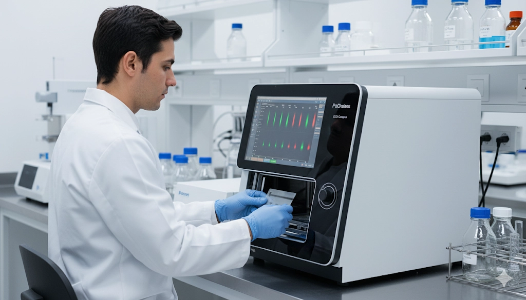
Modern gel imaging systems, equipped with high-resolution, cooled CCD cameras, have largely replaced film. These systems offer several advantages. The digital capture provides a much wider linear dynamic range, allowing for accurate quantification of both high and low-abundance proteins within the same image. The imaging software that accompanies these systems is indispensable for quantitative analysis. It enables precise band-to-band comparisons, calculates band intensity, and provides tools for background subtraction.
For accurate quantification, proper normalization is paramount. This involves standardizing the signal of the target protein to a reference protein, known as a loading control. A loading control is a housekeeping protein, such as GAPDH or actin, that is constitutively expressed and whose expression is not expected to change under the experimental conditions. By dividing the signal of the target protein by the signal of the loading control, researchers can account for differences in protein loading across lanes, ensuring that any observed changes in target protein expression are biologically meaningful and not due to technical variability. This rigorous approach to quantitative analysis is essential for producing reproducible and reliable data in proteomics research.
Best Practices for Western Blotting Data Interpretation and Publication
The final, and arguably most critical, phase of the western blotting workflow is the interpretation of data. The visual inspection of bands on a membrane, while informative, must be supported by a rigorous, quantitative analysis. A critical first step is to confirm that all technical controls are valid. This includes positive controls, which confirm that the antibodies and detection system are working correctly, and negative controls, which verify the specificity of the primary antibody and rule out non-specific binding.
One of the most common pitfalls in western blotting is signal saturation. This occurs when the signal intensity exceeds the linear dynamic range of the detector (either film or a CCD camera), making it impossible to accurately quantify the protein amount. A saturated band appears as a uniform, dark blot and should be considered unreliable for quantitative purposes. Another challenge is the appearance of non-specific bands, which can be minimized by optimizing antibody concentrations and washing steps.
For publication, transparency and reproducibility are key. The scientific community increasingly expects detailed method descriptions, including antibody catalog numbers, dilution factors, and imaging settings. Raw, uncropped images of the entire membrane should be provided to demonstrate the full context of the experiment and to allow for independent verification of the results. By adhering to these best practices, laboratory professionals can enhance the credibility of their research and contribute to a more robust and reliable body of scientific knowledge.
Key Takeaways for Western Blotting
Western blotting remains an indispensable tool in proteomics, enabling the visualization and quantification of proteins with high specificity. The journey from protein sample to publishable data requires a comprehensive understanding of each procedural step, from proper gel electrophoresis and membrane transfer to the selection of advanced immunodetection methods and the use of modern gel imaging systems. By adopting a rigorous, quantitative approach to data analysis and adhering to established best practices for documentation and transparency, researchers can ensure the integrity of their findings and drive meaningful progress in the life sciences. The mastery of these techniques is not merely a technical skill but a commitment to scientific excellence.
Frequently Asked Questions
What is the primary purpose of western blotting in proteomics research?
Western blotting is a key method used to identify and quantify a specific protein of interest within a complex mixture of proteins, providing insights into protein expression.
How is a loading control used in western blotting data analysis?
A loading control, such as a housekeeping protein like actin, is used to normalize the amount of protein loaded into each lane, ensuring accurate and quantitative comparison of protein expression levels.
Why are modern gel imaging systems preferred over X-ray film for western blotting?
Modern CCD camera-based gel imaging systems offer a wider linear dynamic range and greater sensitivity, which allows for more accurate and reliable quantitative analysis of protein expression.
What is multiplexing and how is it achieved in western blotting?
Multiplexing is the simultaneous detection of multiple proteins on a single membrane, typically achieved using fluorescence-based western blotting with secondary antibodies conjugated to different colored dyes.
