Selecting the Best Western Blot Imaging System for Your Lab
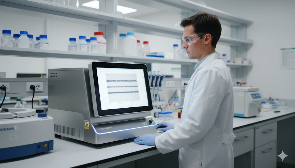
In modern proteomics, the reliability of a laboratory’s research often depends on the fidelity of its data, and few instruments are more central to this process than the western blot imaging system. This critical piece of equipment transforms the molecular signal of a protein into a quantifiable image, allowing for the precise measurement of protein expression and post-translational modifications. A suitable western blot imaging system is essential for obtaining accurate and reproducible results, which are vital for publication and scientific advancement. Navigating the diverse range of available systems requires a careful evaluation of their technical specifications, capabilities, and long-term value. This article provides a comprehensive overview of the key factors to consider when selecting an imaging system to support robust gel imaging and protein analysis.
Understanding Western Blot Imaging System Types: CCD vs. Laser Scanners
The fundamental choice in western blot imaging technology lies between charge-coupled device (CCD) camera-based systems and laser-scanning imagers. Each type offers distinct advantages and is suited for different applications. CCD camera systems are highly versatile and widely used due to their ability to capture a broad spectrum of signals. A single CCD imager can typically detect signals from chemiluminescence, fluorescence, and visible light, making it a flexible platform for various applications, including western blots, DNA gels stained with ethidium bromide, and colorimetric blots. The high sensitivity of cooled CCD cameras allows them to capture the faint, transient light of a chemiluminescent reaction, which is a common detection method for western blotting. Furthermore, they are effective for fluorescence, using LED or filtered light sources to excite fluorophores.
In contrast, laser-scanning imagers are primarily designed for fluorescence-based detection. These systems utilize highly focused lasers to excite specific fluorophores on the membrane, which allows for exceptional signal clarity and resolution. They are particularly well-suited for quantitative multiplexing, where multiple proteins are detected simultaneously on a single blot using antibodies with different fluorescent tags. The use of multiple lasers and filters enables the separation of signals with minimal spectral overlap, leading to highly accurate quantitative data. While laser scanners offer superior performance for certain applications, their utility is often restricted to fluorescence, and they may not be suitable for chemiluminescent or other colorimetric assays without additional hardware. The decision between a CCD system and a laser scanner often comes down to the lab's primary research focus: versatility and broad application versus high-precision, quantitative fluorescence.
Key Technical Specifications for Quantitative Protein Analysis
Selecting a western blot imaging system requires a close examination of several key technical specifications that directly impact data quality. The camera resolution is a critical factor, as it determines the level of detail captured in the image. A higher resolution, measured in megapixels, allows for the precise visualization of closely spaced protein bands. However, resolution must be balanced with pixel size and sensor cooling. The dynamic range of a system is perhaps the most important specification for quantitative analysis. It represents the range of signal intensities the detector can accurately measure, from the faintest detectable signal to the maximum, saturated signal. A wide linear dynamic range is essential for accurately quantifying both high- and low-abundance proteins on a single blot, a common requirement in proteomics research.
Another crucial specification is sensitivity, or the lowest amount of protein that can be reliably detected. High sensitivity is a requirement for detecting proteins expressed at low levels. Finally, the signal-to-noise ratio of the system indicates the clarity of the protein signal relative to background noise. A high signal-to-noise ratio ensures that the detected bands are not obscured by background fluorescence or light, leading to more reliable and publishable data. An ideal system combines high sensitivity with a wide dynamic range and a high signal-to-noise ratio to provide a comprehensive and accurate representation of the proteins on the membrane.
Feature | Low-End System | High-End System |
|---|---|---|
Camera Resolution | ~2 MP | >6 MP |
Dynamic Range | 2-3 orders of magnitude | 4-5 orders of magnitude |
Sensitivity | Nanogram range | Picogram range |
Cooling | Passive | Cooled CCD sensor |
Primary Use | Qualitative, single-band detection | Quantitative, multiplexing |
System Versatility and Workflow Integration
Beyond core imaging technology, the overall functionality and ease of integration into an existing laboratory workflow are significant considerations. A versatile system can save both time and money by accommodating multiple applications. Many modern systems are capable of not only western blotting but also a variety of other gel imaging applications, including nucleic acid gels (e.g., agarose gels with Ethidium Bromide or SYBR Safe), protein gels (e.g., Coomassie Blue or silver stain), and even microplate assays. This multi-application capability can consolidate equipment and simplify laboratory operations.
The software that accompanies the imaging system is equally important. Robust imaging software should offer a user-friendly interface for image acquisition and provide powerful tools for quantitative analysis, such as automated band detection, molecular weight analysis, and background subtraction. Features that facilitate data management, such as the ability to save data in standard file formats and integrate with laboratory information management systems (LIMS), can greatly streamline research. Moreover, ease of use is a critical factor for labs with multiple users. A system with a simple, intuitive workflow and minimal calibration requirements can reduce training time and minimize user-to-user variability, enhancing the reproducibility of results. A well-integrated system with comprehensive software and versatile hardware can serve as a central hub for a lab's gel and protein analysis needs.
Cost and Long-Term Value of Western Blot Imaging Systems
When evaluating a western blot imaging system, the total cost of ownership extends far beyond the initial purchase price. While a lower upfront cost may seem attractive, it is crucial to consider the long-term expenses and potential limitations. The initial investment includes the imager itself, any necessary computer hardware, and the software package. However, ongoing costs can include service contracts, software licenses, and the expense of consumables, such as specific substrates or filters required for a proprietary system. It is important to compare the cost of different detection methods. For example, fluorescence-based detection may have a higher initial hardware cost but can be more cost-effective in the long run by reducing the need for expensive chemiluminescent substrates and saving time with multiplexing.
Furthermore, the longevity and reliability of the equipment are important. A system from a reputable manufacturer with a strong track record of support and regular software updates will provide better value over time. Considering the future needs of the lab is also essential. A system that can grow with the research—for example, by adding new modules or software features—is a more prudent investment than one that may quickly become obsolete. By taking a holistic view of the financial implications, a lab can select a system that not only fits the current budget but also provides a solid foundation for future scientific endeavors.
Making an Informed Decision for Western Blotting
The selection of a western blot imaging system is a significant decision that impacts the quality and efficiency of a laboratory's proteomics research. An informed choice requires a thorough evaluation of the available technologies, from the fundamental differences between CCD cameras and laser scanners to the critical technical specifications that govern data quality. A system’s versatility and integration into the lab’s existing workflow, as well as its total cost of ownership, are also essential factors. By carefully weighing these considerations against the specific needs and goals of the research, a laboratory can invest in a western blot imaging system that provides the robust, quantitative data necessary to drive scientific discovery and innovation. A meticulously chosen system is not merely a tool but a foundational asset for advancing research.
Frequently Asked Questions
How does dynamic range affect western blot quantification?
A wide dynamic range allows for the accurate quantification of both high- and low-abundance proteins within a single image, preventing signal saturation and ensuring reliable data.
What is the difference between a chemiluminescent and a fluorescent western blot system?
A chemiluminescent system detects light produced by a chemical reaction, while a fluorescent system detects light emitted by fluorophores excited by a light source, typically a laser.
Should a lab prioritize resolution or sensitivity when selecting a new system?
Both are important, but for quantitative analysis, a high signal-to-noise ratio and wide dynamic range are often more critical than raw resolution for achieving accurate and reproducible results.
What are the key software features to look for in a modern gel imaging system?
Key software features include automated band detection, precise background subtraction, molecular weight analysis, and tools for data normalization and report generation.
