Key Breakthroughs in Single-Cell and Single-Molecule Microscopy

Written By: Damon Anderson, PhD
Jul 06, 2021
The field of high-resolution microscopy has witnessed several recent breakthroughs.
As reported in a previous post, innovations in image capture and post-image processing have led to significant gains in resolving power.
This overall trend continues with two new exciting developments in the field. The advancements reported below push the limits of resolution even further than before, enabling our ability to view single-molecule interactions and single cell activities in live cells with unprecedented detail.
Barriers to image resolution
Previously, even the highest resolution microscopy techniques could only image structural details of proteins and nucleic acids at levels above nanometer scale. This precluded the ability to map conformational information critical to how these biomolecules interact, signal, and carry out their duties. High resolution structural studies remained in the domain of solid-state techniques, like X-ray crystallography and Cryo-EM, and the large-scale averaging of data involved in these methods.
What is Atomic Force Microscopy and how does it work?
Atomic Force Microscopy (AFM) is a technique that employs a mechanical probe to scan the surface of biological specimens or materials. AFM's tiny but accurate and precise movements can reveal fine details of surface topology at sub-nanometer scales. This technique can achieve atomic resolution of solid surfaces, silicates, semiconductors, etc. However, it cannot achieve this level of resolution for biomolecules in solution or under physiological conditions.
Super-resolution microscopy meets AFM
The authors of a recent study published in Nature borrow a concept from modern optical microscopy to bring higher resolution to AFM imaging. In super-resolution optical microscopy, two fluorescent probes in a sample in very close proximity are visualized with the help of advanced computational algorithms. This in turn permits reconstruction of miniscule measurements between these probes. The investigators of the new technique, termed “Localization Atomic Force Microscopy” (LAFM), harness attributes of both optical and AFM techniques.
Single molecule structures in biological context
As opposed to solid state structural techniques such as X-ray crystallography, which average data from many molecules, the new LAFM technique permits capture of multiple images of single molecules. This provides imaging of atomic level resolution of single molecules in solution and under biological conditions.
A quantum trick
A second recent development focuses on the major issue of resolving single cells using optical microscopy, and specifically the extreme intensity of the light that is required. Microscopes that are used to examine the details of biological systems employ one or two intense light sources. The more powerful the light from these microscopes, the greater the biological detail. However, this comes at the expense of potentially harming or destroying the living specimen.
The authors of a new study published in Nature use a quantum trick to squeeze or condition the light from the photon source. By sending the light beam through a special crystal, only paired photons (quantum pairing) with similar energies are focused on the sample. Removing excess light and noise through this method results in lower intensities and subsequently less damage to biological samples.
Imaging single cells in greater detail
The implication of this new technique includes: greater diagnostic power in determining cancerous from benign cells, better understanding of cell-to-cell communications, greater ability to visualize live-cell events such as gene expression and apoptosis, and many other applications.
Outlook
The advancements described above serve to once again push the resolution barrier, enabling new insights at the atomic level, within single cells, and in the context of living biological processes. Pretty amazing stuff with more that’s sure to come in this intense and innovative field.
View Microscope and Microscope Accessories listings on LabX.com.
Visit the Microscopy and Imaging application page for further resources and product listings.
Feb 12, 2026
Product News
$5M+ in GMP Biopharma Manufacturing & QC Equipment Heads to Online Auction
A Southern California GMP manufacturing and clinical-stage QC laboratory is liquidating new and late-model equipment and unused inventory in an online auction opening February 11, 2026

Feb 12, 2026
Product News
2026 SLAS: Inspiration Catalyzing Discovery
A LabX look at the annual conference and exhibition, a key forum for networking, collaboration, and advancement of new technologies

Feb 11, 2026
Product News
Angstrom Scientific Named U.S. Distributor for Radalytica’s RadalyX Robotic X-ray and CT System
Angstrom Scientific, known for representing microscopy and materials analysis technologies across North America, adds Radalytica’s robotic X-ray and CT platform to its portfolio of analytical and imaging solutions.
Feb 10, 2026
Technical Insight
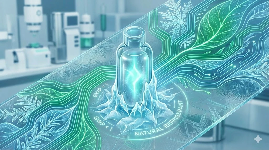
Eco-Friendly Refrigeration: Evaluating Natural Refrigerants and Low-GWP Cooling Systems
This guide explains the operational and environmental benefits of adopting natural refrigerants and low-GWP cooling systems in laboratory settings

Feb 10, 2026
Technical Insight
The Ultimate Guide to Sustainability in Laboratory Equipment and Operations
A comprehensive analysis of sustainable laboratory practices, focusing on energy-efficient equipment, waste management, and digital workflows

Jan 09, 2026
Product News
BRANDTECH Scientific Introduces the Transferpette® pro Micropipette: A New Twist on Comfort and Control
The latest addition to its trusted line of precision liquid handling instruments, engineered for comfort, control, and repeatable accuracy.

Jan 06, 2026
Featured, Technical Insight
New Research Frontiers Unlocked by Next-Generation Flow Cytometry
Next-generation flow cytometry systems using acoustic focusing are opening entirely new areas of research that were previously impractical or inaccessible with conventional flow cytometers.

Jan 05, 2026
Buying Guides
The Bottom Line: How Smart Microcentrifuge Tube Choices Impact Lab Efficiency and Cost
Innovative microcentrifuge tube solutions are more than just lab consumables—they’re strategic tools that can reduce costs, improve outcomes, and support long-term sustainability goals.

Jan 05, 2026
Buying Guides
Future-Proofing Your Lab with Innovative Microcentrifuge Tube Solutions
As laboratories evolve to meet the growing demands of modern science, even the most basic tools—like microcentrifuge tubes—are being reimagined.

Jan 05, 2026
Buying Guides
Microcentrifuge Tube Innovations: Driving Sustainable Change
Integrating next-generation consumables into standard workflows can lead to a more sustainable and responsible research environment.