Microscopy and Imaging Developments in the Life Sciences Universe

Written By: Damon Anderson, PhD
Nov 02, 2021
The life sciences universe is expanding, with several new microscopy and imaging developments pushing the boundaries of our understanding.
Microscopy has long been an area of development and innovation, from early beginnings of the light microscope to the advent of electron microscopes, cutting-edge techniques, and beyond. This growth has included the implementation of microscopy technologies into devices suitable for microplate tissue and cell imaging, high-throughput analysis, and a variety of other applications. Now, several recent developments have pushed the microscopy and imaging fields into new realms of space and time.
Advancing the imaging resolution revolution
We previously reported on ground breaking developments that were forging the path of the imaging resolution evolution. We spoke about imaging resolution and the fact that earlier technologies were incapable of breaking the limits necessary to achieve atomic level structural details of biological molecules and complexes. Several studies and a host of new technologies demonstrated that indeed science was well on its way towards this new super-resolution goal.
Improving 3D imaging accuracy and precision
Fast forward to more recent developments, where we reported on innovations in ultra-high resolution optical microscopy and single-cell (and single-molecule) microscopy. Several key developments included:
- Optical microscopy resolution can be significantly improved using rapid deconvolution of multiple images, and aided by the use of computational deep learning techniques.
- Optical measurements of 1-20 nm can be accomplished using single-molecule localization microscopy (SMLM) techniques combined with the use of advanced probes.
- Single molecules can be imaged in biological contexts by the use of optical microscopy combined with atomic force microscopy, in a technique called Localization Atomic-Force Microscopy (LAFM).
- Single molecules can be imaged in greater detail using a quantum pairing techniques, focusing conditioned photon beams while filtering out damaging excess noise.
What do all of these developments mean for the life sciences?
- Atomic level structural studies of proteins and biomolecular complexes can now be attained, without the inherent problems of solid-state techniques such as x-ray crystallography.
- Single-cell and single-molecule imaging in biological samples is now a reality.
- Advancements in imaging resolution, spatial location, and contrast are creating a richer, more complete view of the life sciences universe.
Tracking rapid biological processes over time
Fast forward to the present, and a new dimension for exploration. Researchers at the ImmunoSensation2 Cluster of Excellence at the University of Bonn have developed techniques that use multi-focal images to reconstruct the movement of fast biological processes in 3 dimensions. This work titled “Multifocal imaging for precise, label-free tracking of fast biological processes in 3D” has been published in Nature Communications.
Many biological processes are rapid, happening on the millisecond time scale. Image acquisition at high frame rates has been used in the past to record fast biological processes. In order to keep track of targets, however, a large field of view and bright fluorescent labels were required. Challenges in maintaining accuracy and precision of watched targets have made previous work tenuous.
The investigators in the present study, focused on the dynamics of flagellar beating in connection with the swimming action of sperm. Using a 3D reconstruction algorithm and multi-focal images, they tracked the movement of non-labeled spherical and filamentous structures quickly and easily, over long time periods and distances. The also charted the fluid flow in the medium surrounding the sperm. How fast and precise were these measurements? The investigators characterized fluid flow and flagellar beating with a z-precision of 0.15 µm, in a volume of 250x260x21 µm, and at high speed of 500 Hz - 2.5 faster sampling speeds than previous work.
Implications of advanced microscopy and imaging technologies
As the authors state, “Life happens in three dimensions”. Nearly all biological materials (cells, organelles, molecular structures) undergo some form of 3D movement. Many of these events occur on the micrometer scale within milliseconds. State-of-the-art microscopy and imaging technologies have made significant impacts on the components of 3D space. Now, as evidenced by this study, the fourth dimension - time - is being explored in even greater accuracy and detail than before.
Tracking rapid biological 3D movements in time will provide the opportunity to better understand biological processes, how they happen and what happens when they go awry. Beyond that, better clarity of these processes may contribute to biomechanical engineering efforts and the development of artificial mechanical systems.
In the current study, the investigators go on to apply their techniques to the actions of insect wings (Drosophila melanogaster), foraging Hydra vulgaris, and crawling Amoeba proteus, to demonstrate the applicability of the approach to a wide variety of movement processes. Such work on an expanding set of subjects may yield valuable insights into how to influence biological processes or replicate these motions for use in our world.
Visit the LabX Microscopy and Imaging application page for further insight and product listings.
Dec 15, 2025
Featured, Popular Products
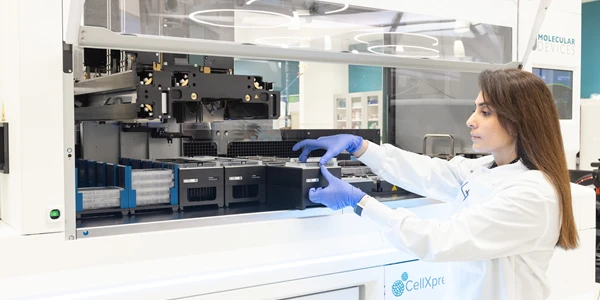
Automated 3D Cell Culture Workflows: Reshaping Disease Research and Drug Development
As 3D cell systems move from exploratory research into routine use across drug development and translational science, automation is becoming essential.
Dec 11, 2025
Technical Insight
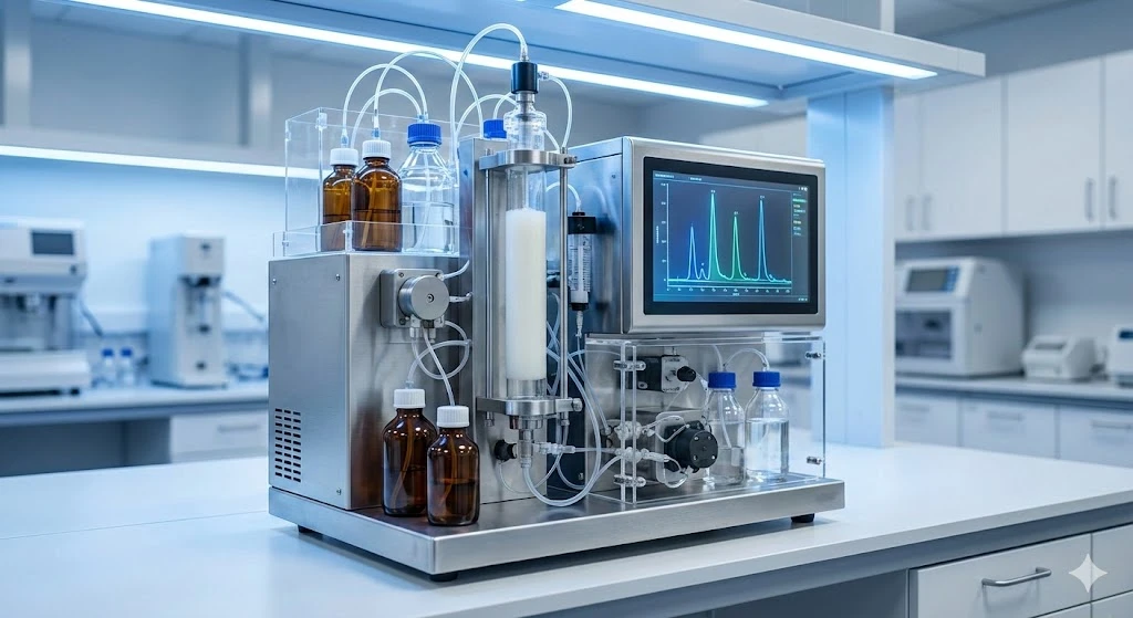
The Best FPLC Systems of 2026
Discover the top FPLC systems of 2026 for protein purification. This guide reviews price, specs, and performance for leading lab equipment models.
Dec 11, 2025
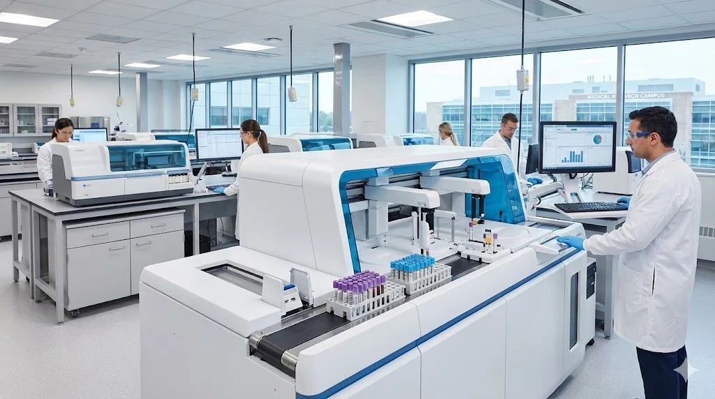
The Best ELISA Kits and Immunoassay Systems of 2026
Discover the top-rated ELISA kits and immunoassay systems of 2026. We review high-throughput analyzers and budget-friendly models for modern labs.
Dec 10, 2025
Featured, Popular Products
How Microplate Dispensers Enable Modern Research and Discovery
From drug discovery to genomics, almost every lab today depends on microplate dispensers.
Dec 08, 2025
Technical Insight
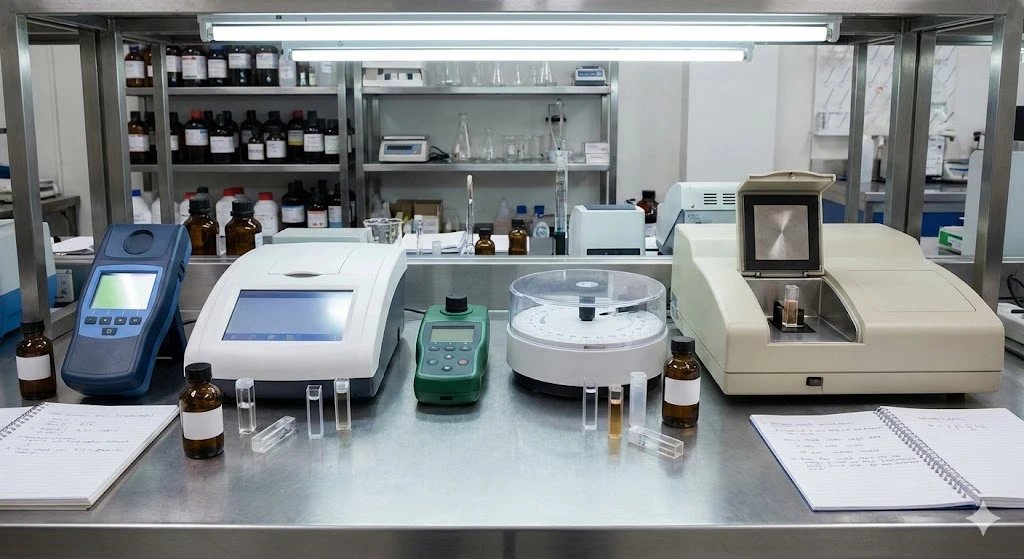
The Best Colorimeters of 2026
Discover the top-rated colorimeters of 2026 for laboratory professionals. We review the best new models for accuracy, speed, and chemical analysis.

Dec 08, 2025
Technical Insight
The Best DNA Sequencers of 2026
Discover the top-performing DNA sequencers of 2026. This guide reviews the latest high-throughput, benchtop, and long-read models for modern laboratories.
Dec 08, 2025
Technical Insight
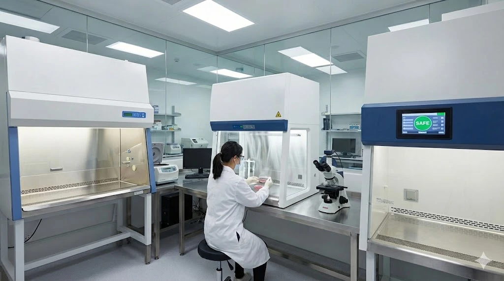
The Best Biosafety Cabinets and Laminar Flow Hoods of 2026
Discover the top biosafety cabinets and laminar flow hoods of 2026. This guide reviews safety, efficiency, and features for optimal laboratory protection.
Dec 05, 2025
Technical Insight
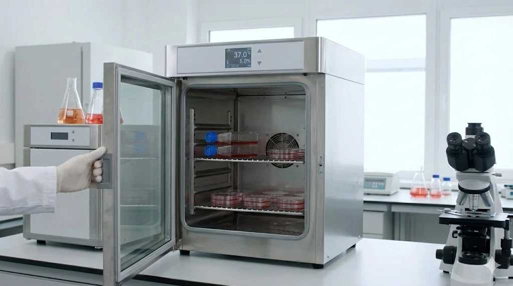
The Best Laboratory Incubators of 2026
Discover the best laboratory incubators of 2026. This guide reviews top models for cell culture and microbiology, focusing on precision, capacity, and new releases.
Dec 05, 2025
Technical Insight
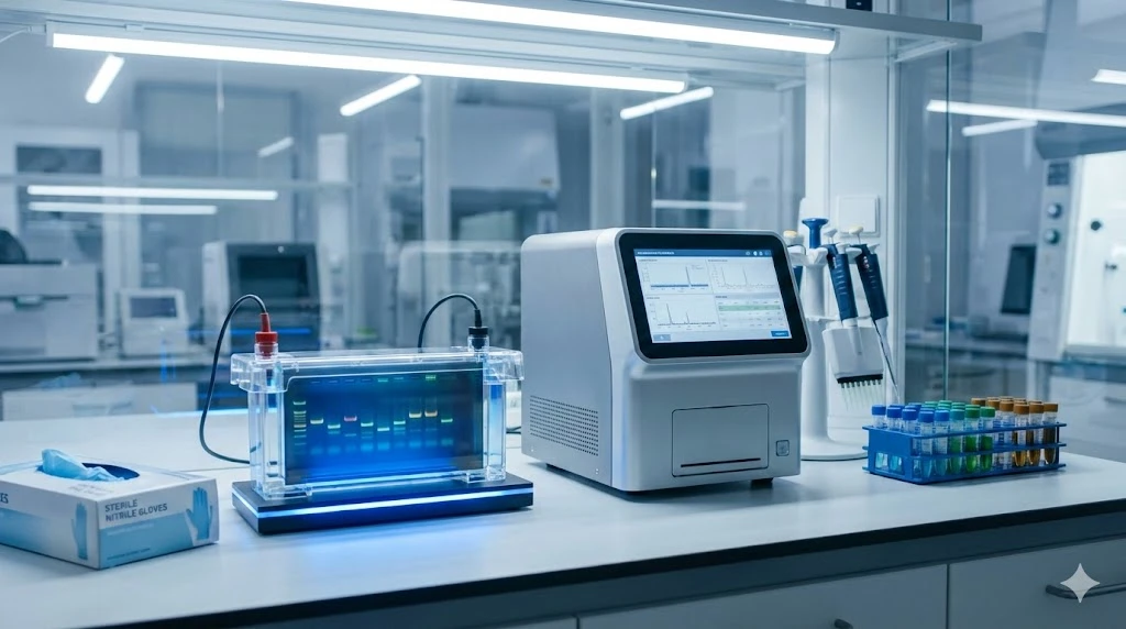
The Best Electrophoresis Equipment of 2026
Discover the top electrophoresis equipment of 2026 for research and clinical labs. Compare models for budget, throughput, and versatility.
Dec 05, 2025
Technical Insight
The Best Laboratory Chillers of 2026
Discover the top-rated cooling systems for 2026. This guide reviews high-performance, eco-friendly, and budget-conscious lab chiller models.