Patch-Clamp Techniques: Choosing Equipment and Optimizing Experiments

Written By: Craig Bradley
Oct 15, 2025
GEMINI (2025)
The field of cellular electrophysiology provides an unparalleled view into the fundamental mechanisms governing excitable cells, yet precise execution of the patch-clamp technique remains a critical challenge. The ability to record currents and potentials from single ion channels or whole cells is foundational for contemporary neuroscience and pharmacology. Successfully conducting high-fidelity patch-clamp experiments requires a nuanced understanding of instrumentation and meticulous protocol optimization. Achieving reliable gigaohm seals and maintaining stable recordings are cornerstones of quantitative electrophysiology. This article details the essential considerations for equipment selection and experimental refinement, crucial steps for any laboratory professional dedicated to advancing electrophysiology research.
Amplifier Selection and Signal Conditioning in Cellular Electrophysiology
The patch-clamp amplifier is the central electronic component in any electrophysiology rig. Its primary function is to convert the minute electrical signal generated across the patch pipette's membrane seal into a measurable voltage output. Selection criteria for this device must prioritize ultra-low-noise operation and versatile compensation features, as the amplifier dictates the noise floor of the entire electrophysiology system. Amplifier headstages, typically integrated with the main unit, contain the current-to-voltage converter and/or a high-input impedance buffer, depending on the mode (voltage-clamp or current-clamp), both critical for accurate single-channel electrophysiology recordings.
Modern amplifiers offer critical capabilities essential for artifact management, notably capacitance neutralization and series resistance compensation. Cell membrane capacitance introduces transient current artifacts that obscure the underlying biological signal. Capacitance neutralization circuitry effectively counteracts these transients, which is particularly important in whole-cell electrophysiology where the cell surface area is large.
Series resistance, originating primarily from the resistance of the patch pipette tip and the access resistance to the cell interior, causes voltage errors when current is flowing. Effective series resistance compensation is mandatory for accurate voltage clamping and precise determination of channel conductance, although it must be applied judiciously to prevent oscillatory feedback that compromises the integrity of the electrophysiology trace.
Furthermore, a well-chosen amplifier must seamlessly transition between voltage-clamp and current-clamp modes. Voltage clamp is the default for studying ion channel kinetics, holding the membrane potential constant while recording ionic currents. Current clamp, conversely, allows for the study of action potential firing and resting membrane potential, where injected current elicits resulting voltage changes. Ensuring the amplifier provides reliable, stable performance in both modes is paramount for versatile electrophysiology investigations.
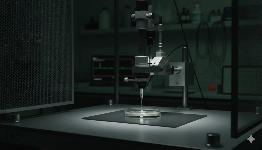
Mechanical Stability: Precision Micromanipulation and Vibration Isolation for Reliable Electrophysiology
The physical stability and precision of the experimental setup are equally as critical as the electronic components in high-quality electrophysiology recordings. The micromanipulator facilitates the controlled movement of the patch pipette toward the cell membrane, demanding nanometer-scale precision for successful seal formation. Two main categories exist: hydraulic/mechanical manipulators and motorized/piezoelectric manipulators. While mechanical systems offer excellent stability and tactile feedback, motorized systems provide digital control, allowing for highly repeatable, programmed movements preferred in automated or high-throughput electrophysiology. The choice is determined by the experimental context; for delicate in vivo patch-clamping, finer, more stable movement control is generally favored.
Vibration isolation is a non-negotiable requirement for patch-clamp electrophysiology. External mechanical noise, even subtle low-frequency vibrations, can disrupt the fragile gigaohm seal. An air-table or specialized passive/active vibration isolation platform is necessary to decouple the entire rig from environmental vibrations. The entire apparatus—including the microscope, stage, and micromanipulators—must rest firmly on this isolated platform.
Vibration isolation (air/active tables) handles mechanical and acoustic disturbances that destabilize seals. A Faraday cage separately shields the preparation from electromagnetic interference. Proper grounding of all electrical components further minimizes baseline noise and system instability.
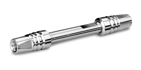
Pipette Fabrication and Internal Solution Chemistry in Electrophysiology Protocols
The patch pipette acts as the primary interface between the electronic measurement system and the biological specimen, making its fabrication a critical skill in electrophysiology. Pipettes are typically pulled from borosilicate or quartz glass capillaries using specialized heated pullers. The resulting taper angle and tip diameter determine the pipette resistance.
Whole-Cell Configuration: Low resistance pipettes (2–5 MΩ) are used to minimize series resistance and facilitate membrane rupture for accessing the cell interior.
Single-Channel Electrophysiology: Higher resistance pipettes (5–10 MΩ) are preferred for single-channel recordings, as they facilitate the formation of high-resistance seals and effectively reduce pipette tip noise.
Reproducible pipette pulling is essential for minimizing variability across electrophysiology experiments.
The composition of the internal (pipette) solution is equally critical, as it controls the intracellular ionic environment once the whole-cell configuration is established through dialysis. Key chemical parameters must be rigorously controlled:
Osmolarity: The solution must closely match the external bath solution (typically 280–310 mOsm for mammalian cells) to prevent osmotic stress, which compromises cell health and seal formation.
pH Buffering: Standard buffers like HEPES or Tris (often adjusted to pH 7.2–7.4) are used to maintain physiological pH for optimal ion channel function.
Ionic Composition: Tailor major ions (e.g., K⁺, Cs⁺, Cl⁻) to the channels under study: use K⁺-based internals for K⁺-current recordings, and Cs⁺-based internals (often with TEA) to suppress K⁺ currents when isolating Na⁺ or Ca²⁺ currents.
Co-factors: Inclusion of ATP and GTP is often necessary in internal solutions to maintain cell viability and metabolic function, as these cofactors are essential for the regulation and modulation of many ion channels, particularly in long-duration recordings.
Meticulous attention to solution preparation, including filtration and accurate concentration measurement, is crucial for successful electrophysiology outcomes and the reliable interpretation of results.
Optimizing Gigaohm Seal Formation and Patch-Clamp Configuration Management
The formation of a Gigaohm (GΩ) seal—an electrical connection between the patch pipette and the cell membrane with resistance typically greater than 1×10⁹Ω—is the defining technical achievement of the patch-clamp technique and is essential for ultra-low-noise electrophysiology. This high seal resistance significantly minimizes current leakage, ensuring that the measured current flows almost exclusively through ion channels.
The sealing process relies on careful pressure management and movement:
| Stage | Action | Electrical Result | Purpose in Electrophysiology |
|---|---|---|---|
| Approach | Gentle movement toward cell; positive pressure applied inside pipette. | Baseline pipette resistance (MΩ). | Keeps the pipette tip clean from debris. |
| Contact | Pipette touches the cell membrane. | Small, immediate increase in resistance. | Confirms physical contact with the cell surface. |
| Seal Formation | Pressure released; mild, continuous negative pressure (suction) applied. | Sharp rise in resistance to >1 GΩ. | Forms the high-resistance electrical seal, eliminating background noise for single-channel electrophysiology. |
Once the GΩ seal is established in the "cell-attached" configuration, the membrane potential remains undisturbed, allowing for single-channel recordings. For whole-cell electrophysiology, a brief, strong pulse of suction is applied to rupture the small membrane patch under the pipette tip, achieving electrical and chemical access to the cell interior.
Other configurations, such as inside-out or outside-out patches, are achieved through subsequent pipette withdrawal maneuvers, allowing for precise pharmacological studies where the internal or external face of the ion channel can be directly accessed and perfused. The technical difficulty lies in maintaining the integrity of the GΩ seal throughout these transitions, a requirement for any successful electrophysiology experiment.
Data Acquisition, Filtering, and Analysis Protocols in Electrophysiology
The final stage of the patch-clamp technique involves the accurate digitization and analysis of the analog current and voltage signals produced by the amplifier. The acquisition hardware (digitizer) must possess sufficient resolution and speed to capture the rapid kinetics inherent in many ion channel processes. A minimum sampling rate of five to ten times the highest frequency component of interest is necessary, although much higher rates are generally preferred to avoid the introduction of aliasing artifacts into the electrophysiology data.
Signal filtering is essential in electrophysiology to improve the signal-to-noise ratio. The analog signal is typically passed through a low-pass Bessel or Butterworth filter to remove high-frequency noise components that lie outside the biologically relevant bandwidth before the signal is digitized. It is crucial to set the filter cutoff frequency (fc) so that the sampling rate is at least five times higher (preferably 10x for single-channel work), minimizing aliasing and preserving kinetic fidelity.
Data analysis relies on specialized software packages designed for electrophysiology. These programs facilitate several critical steps necessary to extract meaningful biological information:
Baseline Correction and Leak Subtraction: These procedures are essential for isolating the current flowing strictly through the ion channels from the background electrical leakage current across the seal.
Kinetic Analysis: This involves fitting the rise and decay phases of current traces to mathematical models (e.g., exponential functions) to derive parameters such as activation and inactivation time constants.
Current-Voltage (I-V) Relationship Studies: Analyzing current-voltage relationships is necessary to determine key biophysical properties, including channel reversal potentials and conductance states.
Single-Channel Analysis (for single-channel electrophysiology): Performing amplitude histograms and dwell-time analyses to characterize unitary conductance and channel gating kinetics.
A rigorous, standardized approach to data acquisition settings and analysis protocols is vital for ensuring the reproducibility and scientific validity of all electrophysiology studies.
Advancing Precision in Cellular Electrophysiology
The pursuit of high-resolution electrophysiology depends equally on sophisticated instrumentation and the meticulous adherence to rigorous experimental protocols. Successfully executing patch-clamp techniques requires the careful selection of ultra-low-noise amplifiers and high-precision micromanipulators, all housed within an electromagnetically shielded and vibration-dampened environment. Furthermore, the ability to consistently pull high-quality patch pipettes and meticulously prepare internal solutions directly determines the quality of the gigaohm seal, which is the technical foundation of reliable electrophysiology data acquisition. A standardized approach to signal filtering and specialized software-based analysis completes the pipeline, ensuring that the resulting insights into ion channel function and cellular excitability are both accurate and reproducible, consistently elevating the standard of cellular electrophysiology.
Frequently Asked Questions in Electrophysiology
What is the minimum required resistance for a successful Gigaohm seal?
For reliable patch-clamp electrophysiology, the seal resistance should ideally be 1 GΩ (Gigaohm) or higher. Seals typically in the 1 to 5 GΩ range are considered excellent, significantly reducing background current leakage and ensuring that measured currents are predominantly from ion channels.
How does series resistance compensation affect whole-cell electrophysiology?
Series resistance compensation is crucial because uncompensated resistance introduces voltage errors, causing the actual membrane potential to deviate from the command potential, especially when large currents are flowing. Proper compensation improves voltage control and the accuracy of kinetic measurements in whole-cell electrophysiology.
What is the role of the Faraday cage in patch-clamp experiments?
The Faraday cage shields the sensitive electrophysiology recording area, particularly the headstage and preparation, from external electromagnetic interference. This shielding is essential for reducing high-frequency noise and maintaining the ultra-low-noise environment required for all forms of electrophysiology.
Which parameters are critical when preparing patch pipette solutions?
The most critical parameters are osmolarity, which must match the bath solution to prevent cell damage; pH, which should be maintained at a physiological level (typically 7.2 to 7.4); and ionic composition, which is tailored to the specific ion channels being studied in the electrophysiology experiment.
This article was created with the assistance of Generative AI and has undergone editorial review before publishing.
Mar 02, 2026
Product News
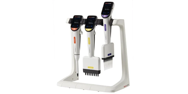
Thermo Fisher Launches Fluid Ease™ Pro ClipTip™ Electronic Pipette with High-Resolution Touchscreen and Advanced Workflow Controls
The system combines programmable control, intelligent power management, and a secure tip attachment mechanism to support consistent, reproducible liquid handling.
Feb 26, 2026
Product News
Waters Launches Microflow LC Columns with MaxPeak Premier Technology for Higher Sensitivity and Lower Resource Consumption
The product launch reflects continued investment in chromatographic chemistries that address real-world bottlenecks in method development and routine analysis
Feb 19, 2026
Technical Insight
Implementing Hydrogen Generators for Clean Lab Gas Supply: Safety and Benefits
Transitioning to on-site gas production streamlines analytical workflows, eliminates hazardous cylinder storage, and supports facility sustainability.

Feb 19, 2026
Technical Insight
Waste Minimization in Laboratory Settings: Best Practices for Chemical, Biological, and Electronic Waste
Effective waste minimization in laboratories improves safety and sustainability. Explore methods for managing chemical, biological, and electronic waste.
Feb 19, 2026
Technical Insight
Circular Economy in Laboratory Equipment: How to Adopt Repair, Reuse, and Refurbishment Strategies
Adopting a circular economy for laboratory equipment minimizes environmental impact while maximizing instrument lifespan and optimizing laboratory budgets.
Feb 12, 2026
Product News
$5M+ in GMP Biopharma Manufacturing & QC Equipment Heads to Online Auction
A Southern California GMP manufacturing and clinical-stage QC laboratory is liquidating new and late-model equipment and unused inventory in an online auction opening February 11, 2026

Feb 12, 2026
Product News
2026 SLAS: Inspiration Catalyzing Discovery
A LabX look at the annual conference and exhibition, a key forum for networking, collaboration, and advancement of new technologies
Feb 11, 2026
Product News
Angstrom Scientific Named U.S. Distributor for Radalytica’s RadalyX Robotic X-ray and CT System
Angstrom Scientific, known for representing microscopy and materials analysis technologies across North America, adds Radalytica’s robotic X-ray and CT platform to its portfolio of analytical and imaging solutions.

Feb 10, 2026
Technical Insight
Eco-Friendly Refrigeration: Evaluating Natural Refrigerants and Low-GWP Cooling Systems
This guide explains the operational and environmental benefits of adopting natural refrigerants and low-GWP cooling systems in laboratory settings
Feb 10, 2026
Technical Insight
The Ultimate Guide to Sustainability in Laboratory Equipment and Operations
A comprehensive analysis of sustainable laboratory practices, focusing on energy-efficient equipment, waste management, and digital workflows