Electrophysiology Systems: Principles, Instrumentation, and Applications in Neuroscience and Cardiology

Written By: Craig Bradley
Oct 15, 2025
GEMINI (2025)
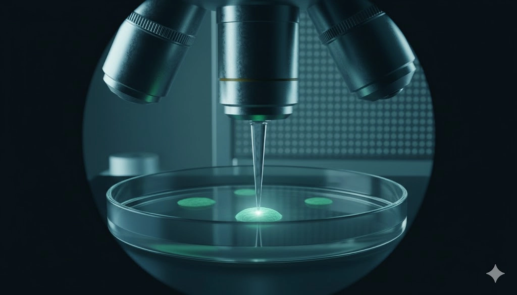
The accurate measurement of electrical activity in living cells is paramount to advancing understanding across numerous biological fields, placing electrophysiology at the core of high-impact laboratory research. Mastery of electrophysiology systems, which measure the ionic current flow that dictates cellular excitability, is indispensable for laboratory professionals seeking robust scientific outcomes and adherence to industry standards, particularly in the pharmaceutical and academic sectors. The methodologies employed in this discipline, ranging from high-resolution single-cell recordings to non-invasive, large-scale mapping, offer profound insights into cellular function, disease mechanisms, and therapeutic efficacy, thus becoming a critical workflow component in any laboratory focused on excitable tissues. This comprehensive overview details the fundamental principles, essential instrumentation, and key applications of modern electrophysiology systems across neuroscience and cardiology.
Foundational Principles: Cellular Electrophysiology, Ion Flux, and Membrane Potential
Understanding the electrical phenomena within a cell requires a solid grasp of fundamental membrane biophysics. The basis of cellular electrophysiology is the membrane potential, which is established by the unequal distribution of ions, primarily sodium (Na+), potassium (K+), chloride (Cl-), and calcium (Ca2+), across the cell membrane. This potential difference is maintained by the selective permeability of the lipid bilayer and the action of active ion transporters, such as the Na+/K+-ATPase pump.
Generating and Measuring the Membrane Potential
The resting membrane potential results from the steady-state efflux of K+ ions through leak channels, a process accurately described by the Goldman-Hodgkin-Katz equation, which accounts for the permeability of multiple ions. Perturbations to this state, driven by external stimuli or synaptic input, cause transient changes in ion permeability mediated by voltage-gated or ligand-gated ion channels. These changes manifest as action potentials, the fundamental unit of electrical signaling in neurons and muscle cells.
From a laboratory perspective, the principles of measurement rely on establishing an electrical path between the cell’s interior or surface and a high-impedance amplifier. Key concepts include:
Capacitive Current: The instantaneous current flow required to charge or discharge the membrane capacitance when the voltage is changed. This is a critical factor when interpreting fast transient signals.
Ohm's Law in Electrophysiology: The relationship between voltage (
V ), current (I ), and resistance (R ) is applied to various components, including the pipette resistance, the seal resistance, and the input resistance of the cell membrane. High seal resistance is necessary for minimizing leakage current and maximizing the signal-to-noise ratio in high-resolution recordings.The Nernst Equation: This equation determines the equilibrium potential for a single ion species, providing a theoretical maximum or minimum voltage that the cell can reach based on that ion's concentration gradient. This calculation is vital for selecting appropriate internal and external solutions in an electrophysiology setup.
Accurate data acquisition demands precise control over the ionic environment and temperature, highlighting the dependence of successful laboratory outcomes on meticulous solution preparation and maintenance of physiological conditions.
High-Resolution Electrophysiology: Mastering Patch-Clamp Techniques for Single-Cell Analysis
The patch-clamp technique remains the definitive methodology for high-resolution measurement of ionic currents across biological membranes. Developed in the late 1970s, it revolutionized cellular electrophysiology by allowing the detection of currents flowing through single ion channels, a feat of instrumentation achieved by establishing a high-resistance, gigaohm
Configurations and Their Analytical Applications
The versatility of the patch-clamp setup lies in its various recording configurations, each optimized for specific analytical goals:
Cell-Attached Mode: The initial configuration where the giga-seal is established. It is non-invasive and allows the study of single-channel activity in the native cellular environment, providing invaluable data on channel gating kinetics under physiological conditions.
Inside-Out Patch: Achieved by retracting the pipette from the cell after the cell-attached mode, this configuration exposes the intracellular face of the membrane patch to the bath solution. This is ideal for studying the effects of intracellular messengers (e.g., ATP, kinases) on channel function.
Outside-Out Patch: Created by rapid withdrawal of the pipette after establishing the whole-cell configuration. The patch membrane reseals with the extracellular surface facing outward. This is the preferred configuration for characterizing the response of ligand-gated channels to neurotransmitters or pharmaceutical agents delivered via the bath.
Whole-Cell Mode: This is the most common configuration for measuring macroscopic currents. A brief suction pulse ruptures the small patch of membrane under the pipette tip, allowing low-resistance electrical access to the entire cell interior. This method is used to record overall membrane properties, such as action potentials and synaptic currents, and is widely utilized in drug screening assays, providing crucial data on a compound's effect on total cellular excitability.
The rigorous setup for patch-clamp requires a high-impedance amplifier, an inverted microscope with differential interference contrast (DIC) optics for visualization, and a vibration-isolation table to mitigate mechanical noise, which could otherwise mask the picoampere-level single-channel currents. The precision and resolution of patch-clamp data set the benchmark for studying ion channel function.
High-Throughput Electrophysiology: Multi-Electrode Arrays (MEAs) for Drug Screening
While patch-clamp offers high temporal and spatial resolution for single-cell analysis, many research objectives, particularly in high-throughput screening and network-level analysis, demand the capacity to record simultaneously from hundreds or thousands of cells. Multi-electrode arrays (MEAs) address this need, representing a significant advancement in the capabilities of modern electrophysiology systems.
MEA Methodology and Advantages
MEAs consist of a substrate (typically glass or plastic) featuring a grid of embedded microelectrodes, ranging from 8 to over 4,096 in number, to which excitable cells (e.g., primary neurons, induced pluripotent stem cell (iPSC)-derived cardiomyocytes) are cultured. The electrodes are non-invasively coupled to the cell membrane, allowing the recording of extracellular field potentials (FPs) generated by propagating action potentials across the cell population or network.
The core advantages of employing multi-electrode arrays in the laboratory setting include:
Non-Invasiveness and Longevity: Recordings are made extracellularly, preserving the integrity of the cells and allowing for long-term monitoring (weeks to months) of network development, maturation, and response to chronic drug exposure.
Network-Level Analysis: MEAs capture the synchronous firing patterns and connectivity of complex biological networks, providing data on neural burst activity, synchronization indices, and conduction velocity, metrics that are unattainable with single-cell techniques.
High-Throughput Potential: The format is easily adaptable to 96-well and 384-well plate formats, making MEAs a powerful tool in preclinical drug development. They enable rapid screening of compound libraries to identify potential neurotoxicology or proarrhythmic risk, significantly accelerating laboratory workflows.
Feature | Patch-Clamp | Multi-Electrode Array (MEA) |
|---|---|---|
Resolution | Single-channel, whole-cell (picoampere) | Extracellular field potential (microvolt) |
Cell Count | One cell per recording | Hundreds to thousands (network) |
Invasiveness | Highly invasive (membrane breach) | Non-invasive (extracellular) |
Application Focus | Detailed kinetics, single-channel pharmacology | Network activity, cardiotoxicity screening |
The integration of multi-electrode arrays into automated liquid handling systems has solidified their position as an indispensable platform for translational electrophysiology research, especially in assessing drug safety profiles and disease modeling.
Signal Amplification and Noise Mitigation: Critical Steps in Electrophysiology Data Integrity
Regardless of whether the methodology involves high-impedance patch-clamp or lower-impedance multi-electrode arrays, the integrity of the final recorded signal hinges upon the quality of signal amplification and the effectiveness of noise reduction strategies. Cellular currents are inherently minuscule, often in the nanoampere or picoampere range, necessitating sophisticated electronic circuits to bring the signal into a measurable range while preserving its fidelity.
Instrumentation Principles for High-Fidelity Recording
A typical electrophysiology recording system consists of three main components, all critical for data quality:
The Amplifier (Headstage and Main Amplifier): The headstage, positioned close to the recording electrode, converts the high-impedance voltage signal into a low-impedance voltage signal that can be transmitted to the main amplifier with minimal loss and noise pickup. The main amplifier then provides the necessary gain (often up to 10,000x or more) and performs essential functions like voltage or current clamping.
Filtering: Both analog and digital filtering are applied to remove unwanted frequency components. Low-pass filters are used to eliminate high-frequency noise, which is often inherent in the electronic environment, but care must be taken to ensure the filter cutoff frequency does not distort the biologically relevant high-frequency components of the action potential.
Digitization and Analysis: The amplified and filtered analog signal must be converted to a digital format (ADC) for computer storage and analysis. The sampling rate must adhere to the Nyquist theorem (sampling at least twice the highest frequency of interest) to prevent aliasing and ensure accurate representation of the cellular events.
Noise Mitigation Strategies
Laboratory professionals must proactively manage sources of noise to achieve high-quality electrophysiology data. The primary sources of noise include:
Thermal Noise (Johnson Noise): Generated by the random motion of charge carriers within resistive elements (pipette tip, seal resistance). This noise is unavoidable but can be minimized by lowering the resistance of the recording pipette.
60 Hz Noise (Mains Noise): Electromagnetic interference from power lines and laboratory equipment. This is typically addressed by rigorous shielding (Faraday cages) and the use of grounding and earth loops. A differential amplifier often helps to reject this common-mode noise.
Shot Noise: Associated with the discrete nature of current flow (individual ion channels opening and closing). This is most prevalent in single-channel patch-clamp recordings.
Effective signal amplification and noise control are not merely instrumental considerations; they are the foundation upon which quantitative analysis and valid scientific conclusions in electrophysiology are built.
Advanced Applications: Optogenetics, Neural Circuit Mapping, and Cardiac Arrhythmias Modeling
The sophistication of modern electrophysiology instrumentation has enabled its application to complex biological questions across diverse research areas, most notably in neuroscience and cardiology. The ability to precisely measure electrical activity provides a functional readout of cellular health and communication.
Neuroscience Applications: Optogenetics and Neural Circuits
In neuroscience, the combination of electrophysiology with advanced molecular tools has unlocked the study of defined neural circuits. Optogenetics, a technique utilizing light-sensitive microbial proteins (opsins) genetically expressed in targeted cells, allows for precise, millisecond-scale control over neuronal activity.
The application involves:
Targeted Expression: A specific subset of neurons (e.g., inhibitory interneurons) is engineered to express an opsin, such as Channelrhodopsin-2 (ChR2), which acts as a light-gated ion channel.
Light Stimulation: Focused light, delivered via fiber optics or LEDs, precisely controls the activity of the targeted neurons—either activating them (ChR2) or silencing them (Halorhodopsin, NpHR).
Electrophysiological Readout: Simultaneous patch-clamp or multi-electrode arrays recording measures the resulting electrical changes in the target cells or the downstream circuit.
This synergistic approach provides the temporal precision of light-mediated control with the functional resolution of electrophysiology, enabling laboratory professionals to map neural connectivity and determine the functional roles of specific cell types within complex circuits in vivo and in vitro.
Cardiology Applications: Modeling Cardiac Arrhythmias
In cardiology, electrophysiology is critical for assessing the safety and efficacy of novel therapeutic agents and for understanding disease pathology. The measurement of action potentials and spontaneous beating patterns in human iPSC-derived cardiomyocytes serves as a gold standard for predicting cardiotoxicity and for modeling cardiac arrhythmias.
Drug-Induced Proarrhythmia: Compounds that block the
hERG potassium channel are known to prolong the cardiac action potential, which can lead to a potentially fatal form of cardiac arrhythmias known as Torsades de Pointes. Multi-electrode arrays are particularly well-suited for screening this risk, as they can monitor changes in the field potential duration (FPD), which correlates with the action potential duration (APD).Disease Modeling: iPSC technology allows researchers to generate cardiomyocytes from patients with inherited arrhythmia syndromes. Recording the electrical phenotype of these disease models via patch-clamp (for detailed single-cell analysis) or MEA (for network-level conduction defects) provides mechanistic insights into the genesis of cardiac arrhythmias and offers a platform for personalized medicine.
The integration of these diverse methodologies underscores the centrality of electrophysiology in modern biomedical discovery, bridging basic science and translational medicine.
Achieving Professional Excellence in Electrophysiology Research
The continued evolution of electrophysiology systems presents laboratory professionals with powerful, high-resolution tools for functional analysis of excitable cells. Achieving mastery in this domain requires not only an understanding of the fundamental principles of membrane biophysics and ion flux but also a command of the intricacies of instrumentation, including advanced signal amplification and noise rejection techniques. The transition from manual, low-throughput methods like patch-clamp to automated, high-content platforms using multi-electrode arrays allows for scalable discovery efforts, while the integration of molecular tools like optogenetics provides unprecedented experimental control in neuroscience. Professionals capable of navigating this diverse landscape, applying the correct methodology for recording action potentials, characterizing ion channel function, and modeling conditions such as cardiac arrhythmias, are positioned to generate the rigorous, quantitative data necessary for significant advances in drug development and disease understanding.
Frequently Asked Questions (FAQ)
What defines a successful Giga-seal in patch-clamp electrophysiology, and why is it critical?
A successful Giga-seal in patch-clamp electrophysiology is defined as an electrical resistance greater than one Gigaohm
How do multi-electrode arrays (MEAs) accelerate cardiotoxicity screening compared to traditional methods?
Multi-electrode arrays significantly accelerate cardiotoxicity screening by providing a non-invasive, high-throughput platform for functional electrophysiology measurements. Instead of relying on manual, single-cell patch-clamp recordings, MEAs allow simultaneous and continuous recording of extracellular field potentials from hundreds of cardiomyocytes across multiple wells. This enables rapid quantification of key cardiotoxicity metrics, such as beat rate, beat regularity, and field potential duration (FPD), which directly correlate with the risk of cardiac arrhythmias. The MEA system's ability to monitor these parameters over hours or days, automatically and with high reproducibility, dramatically reduces the labor and time required to assess the proarrhythmic potential of drug candidates, making it a cornerstone of contemporary preclinical safety assessment.
What is the advantage of combining optogenetics with traditional electrophysiology recording?
The primary advantage of integrating optogenetics into traditional electrophysiology is the unparalleled spatiotemporal precision it brings to controlling and studying excitable cells. While electrophysiology provides the functional readout of cellular activity, optogenetics provides a mechanism to manipulate that activity using light, with millisecond resolution. This combination allows researchers using techniques like patch-clamp or multi-electrode arrays to precisely activate or inhibit genetically defined cell populations, isolating the role of specific neural circuits or cell types. This capability is crucial for accurately mapping connectivity, determining causal relationships in neural signaling, and dissecting the function of complex circuits that underpin neurological disorders, thereby generating highly controlled and mechanistically informative electrophysiology data.
What are the main sources of noise that laboratory professionals must mitigate in an electrophysiology system?
Laboratory professionals must proactively mitigate several key sources of noise in an electrophysiology system to ensure successful signal amplification and data fidelity. The three main categories are thermal noise, shot noise, and environmental noise. Thermal noise, which arises from the random motion of charge carriers in the pipette and seal resistance, is intrinsic and proportional to temperature. Shot noise is related to the discrete nature of current flow through channels, primarily affecting single-channel patch-clamp recordings. Environmental noise, chiefly
This article was created with the assistance of Generative AI and has undergone editorial review before publishing.
Mar 02, 2026
Product News
Thermo Fisher Launches Fluid Ease™ Pro ClipTip™ Electronic Pipette with High-Resolution Touchscreen and Advanced Workflow Controls
The system combines programmable control, intelligent power management, and a secure tip attachment mechanism to support consistent, reproducible liquid handling.
Feb 26, 2026
Product News
Waters Launches Microflow LC Columns with MaxPeak Premier Technology for Higher Sensitivity and Lower Resource Consumption
The product launch reflects continued investment in chromatographic chemistries that address real-world bottlenecks in method development and routine analysis

Feb 19, 2026
Technical Insight
Implementing Hydrogen Generators for Clean Lab Gas Supply: Safety and Benefits
Transitioning to on-site gas production streamlines analytical workflows, eliminates hazardous cylinder storage, and supports facility sustainability.

Feb 19, 2026
Technical Insight
Waste Minimization in Laboratory Settings: Best Practices for Chemical, Biological, and Electronic Waste
Effective waste minimization in laboratories improves safety and sustainability. Explore methods for managing chemical, biological, and electronic waste.
Feb 19, 2026
Technical Insight
Circular Economy in Laboratory Equipment: How to Adopt Repair, Reuse, and Refurbishment Strategies
Adopting a circular economy for laboratory equipment minimizes environmental impact while maximizing instrument lifespan and optimizing laboratory budgets.
Feb 12, 2026
Product News
$5M+ in GMP Biopharma Manufacturing & QC Equipment Heads to Online Auction
A Southern California GMP manufacturing and clinical-stage QC laboratory is liquidating new and late-model equipment and unused inventory in an online auction opening February 11, 2026

Feb 12, 2026
Product News
2026 SLAS: Inspiration Catalyzing Discovery
A LabX look at the annual conference and exhibition, a key forum for networking, collaboration, and advancement of new technologies
Feb 11, 2026
Product News
Angstrom Scientific Named U.S. Distributor for Radalytica’s RadalyX Robotic X-ray and CT System
Angstrom Scientific, known for representing microscopy and materials analysis technologies across North America, adds Radalytica’s robotic X-ray and CT platform to its portfolio of analytical and imaging solutions.

Feb 10, 2026
Technical Insight
Eco-Friendly Refrigeration: Evaluating Natural Refrigerants and Low-GWP Cooling Systems
This guide explains the operational and environmental benefits of adopting natural refrigerants and low-GWP cooling systems in laboratory settings

Feb 10, 2026
Technical Insight
The Ultimate Guide to Sustainability in Laboratory Equipment and Operations
A comprehensive analysis of sustainable laboratory practices, focusing on energy-efficient equipment, waste management, and digital workflows