Introduction to Multi-Electrode Arrays for Neural Network Studies

Written By: Craig Bradley
Oct 15, 2025
GEMINI (2025) The foundational understanding of neural network function, synaptic communication, and pharmacological responses in neuronal cultures is increasingly dependent on high-fidelity electrophysiological recording platforms. The development of multi-electrode arrays represents a paradigm shift in neuroscientific research, enabling long-term, non-invasive monitoring of complex cellular behavior. Laboratory environments focused on drug discovery and disease modeling benefit significantly from the use of multi-electrode arrays as they provide a spatial and temporal map of activity across thousands of neurons. This technology is critical for advancing in vitro models by moving beyond single-cell patch-clamp techniques to capture genuine network-level activity and monitor the integrity of the neural system. A multi-electrode array system is fundamentally defined by its physical hardware, which facilitates intimate and stable coupling between electronic components and biological specimens. The typical architecture of a multi-electrode array consists of a transparent substrate—often glass or polymer—upon which an arrangement of highly conductive microelectrodes is fabricated. These electrodes are typically made from inert materials such as platinum, titanium nitride, or indium tin oxide, selected for their biocompatibility and low impedance characteristics, which are essential for minimizing noise and maximizing signal quality during the recording of neuronal activity. The spatial arrangement of the electrodes is a critical design feature; they are generally distributed in a regular grid pattern, ranging from 8x8 to 256 or more distinct recording sites, providing simultaneous spatial coverage over a large area of the cultured cells. The primary function of the substrate is to anchor the neuronal culture and maintain a sterile, controlled microenvironment. Wells are often etched into the substrate surface to physically constrain the neural population, encouraging the formation of organized, interconnected networks. The dimensions of the microelectrodes themselves—typically ranging from 10 to 30 micrometers in diameter—are optimized to effectively capture the minute extracellular electrical signals generated by adjacent neurons. A central reference electrode is also incorporated into the design of the multi-electrode array to serve as the ground for the recording system, ensuring signal stability. Advances in multi-electrode arrays technology are continuously pushing the boundaries of spatial and temporal resolution. High-density MEAs (HD-MEAs) now incorporate thousands of electrodes packed closely together (with pitches as low as 17 The operational core of multi-electrode arrays is the principle of extracellular field potential recording. Unlike intracellular recording techniques that measure voltage changes across the cell membrane, MEAs detect the small voltage fluctuations—on the order of microvolts—that occur in the extracellular space surrounding the active neurons. These fluctuations are the result of transient ion currents flowing across neuronal membranes during action potential generation (spikes) and synaptic communication (local field potentials). The electrical activity is capacitively coupled to the microelectrodes, amplified by a dedicated multi-electrode array amplifier system, and digitized for processing. The digitized data collected by the multi-electrode array contains two primary forms of information: the action potentials, or "spikes," and the lower-frequency local field potentials (LFPs). Spikes, which represent the primary output signals of individual neurons, manifest as sharp, high-frequency transients. Sophisticated signal processing techniques, collectively termed spike sorting, are applied to this data to distinguish the unique electrical signature of each neuron firing near a given electrode. This process allows for the tracking of individual neuronal units within the dense network. Local Field Potentials, on the other hand, are broad, low-frequency oscillations reflecting the synchronous synaptic input and dendritic processing within a larger population of neurons. Analysis of LFPs provides crucial insight into population-level synchronicity, oscillatory dynamics, and the coordinated behavior of the neuronal network, which is particularly relevant in the study of neurological disorders characterized by altered rhythmicity. The ability of the multi-electrode array system to simultaneously capture both single-unit activity and network-level rhythmicity makes it an unparalleled tool for comprehensive neurophysiological assessment. The utility of multi-electrode arrays extends deeply into translational research, particularly within the domains of drug development, toxicology screening, and the creation of high-fidelity disease models. The scalability and throughput potential of MEA systems surpass traditional manual electrophysiology, allowing laboratories to test hundreds of compounds across multiple culture batches concurrently. In pharmaceutical screening, MEAs offer a functional readout that is highly predictive of drug efficacy and potential neurotoxicity. Compounds are introduced to the cultured networks, and the resulting changes in electrophysiological parameters are monitored in real-time. This provides a direct, physiological measure of compound effect, which can include: Changes in Firing Rate: Assessing excitability or suppression of network activity. Modulation of Bursting Activity: Analyzing alterations in the frequency or duration of high-intensity firing epochs. Synchronicity Analysis: Evaluating the compound’s impact on the coordinated communication between distant network nodes, a key indicator of synaptic function. Long-Term Potentiation (LTP) and Depression (LTD) Screening: Using integrated stimulation protocols to assess compound effects on synaptic plasticity mechanisms, which are crucial for learning and memory research. For modeling complex neurological diseases in vitro, multi-electrode arrays provide the capability to detect subtle phenotypic changes associated with pathology. For instance, in models utilizing induced pluripotent stem cell (iPSC)-derived neurons from patients with autism, schizophrenia, or epilepsy, MEA recordings can reveal abnormal hyperexcitability, reduced network connectivity, or aberrant oscillatory patterns long before structural or biochemical changes become apparent. Epilepsy Modeling: MEAs are invaluable for monitoring the spontaneous development of seizure-like bursts (hypersynchrony) in pro-epileptic cultures and for testing anticonvulsant drugs. Neurodegenerative Disorders: In models of Alzheimer’s and Parkinson’s disease, MEA analysis can detect early signs of network silencing or disorganized activity, providing functional endpoints for therapeutic intervention studies. The standardization and adoption of multi-electrode arrays in regulated environments are making MEA data increasingly accepted as a robust, non-subjective measure of neural function. The sheer volume and complexity of data generated by a single multi-electrode array experiment necessitate the use of specialized analytical software and advanced algorithms. A high-density MEA recording for one minute can yield gigabytes of raw voltage traces across hundreds of channels, making manual inspection infeasible. Effective interpretation requires transforming this raw data into quantifiable, biologically meaningful metrics. Key metrics used for characterizing network function derived from multi-electrode arrays include: Metric Category Specific Measurement Biological Significance Single-Unit Activity Mean Firing Rate (MFR) Baseline neuronal excitability and viability. Temporal Dynamics Inter-Spike Interval (ISI) Distribution Characterization of firing patterns (e.g., random vs. regular). Network-Level Metrics Burst Rate, Burst Duration, Spikes per Burst Measurement of the intensity and stability of synchronized firing events. Connectivity/Synchrony Network Spikes Synchrony Index (NSSI) Quantification of the degree of coordinated activity across the entire network. Propagation Cross-correlation, Time-Lag Analysis Determination of directional communication and functional pathways within the culture. Software systems perform spike detection, spike sorting, and subsequent feature extraction. For spike sorting, algorithms are used to classify detected voltage peaks based on shape and amplitude, isolating the contributions of different neurons. Following sorting, data visualization tools are used to generate raster plots—graphical representations of the timing of every detected spike across all channels—which visually reveal the synchronicity and bursting behavior of the neural population. Challenges in data interpretation often center on distinguishing true biological effects from artifacts or normal biological variability. Robust statistical methods and rigorous quality control steps are applied to ensure that changes in metrics like MFR or burst rate are statistically significant and reflect the intended manipulation (e.g., drug exposure or disease progression) rather than mere culture drift or noise from the multi-electrode array equipment. The proper use of appropriate controls and standardized analysis pipelines is crucial for generating reproducible results from multi-electrode arrays. The field of multi-electrode arrays is rapidly evolving, with future advancements centered on increasing integration, improving fidelity, and expanding applicability. The development of three-dimensional (3D) multi-electrode arrays and systems capable of interfacing with organoids is a major area of growth, moving beyond 2D planar cultures to capture more complex, in vivo-like tissue architectures and organization. Further innovation includes the integration of microfluidic systems directly onto the MEA platform. This combination allows for precise control over the cellular microenvironment, enabling complex perfusion, rapid drug application, and the creation of compartmentalized cultures that mimic physiological barriers. The continuous refinement of electrode materials, particularly the shift toward carbon nanotube and graphene-based electrodes, promises ultra-low noise recordings and enhanced long-term stability. As analytical tools leverage machine learning and deep learning algorithms, the interpretation of complex MEA data, especially in identifying novel disease biomarkers, will become increasingly automated and accurate, further solidifying the multi-electrode array as an indispensable technology in neurobiology laboratories. What is the primary advantage of using multi-electrode arrays over patch-clamp techniques?
Multi-electrode arrays (MEAs) offer a high-throughput, non-invasive method for monitoring the electrophysiological activity of entire neural networks simultaneously over extended periods (weeks to months). In contrast, patch-clamp is an invasive, high-resolution technique typically limited to measuring the activity of one or a few cells over a short duration. How do multi-electrode arrays differentiate between action potentials and local field potentials?
The distinction is based on the signal frequency. Action potentials (spikes), representing individual neuronal firing, are typically high-frequency signals (above 100 Hz). Local field potentials (LFPs), reflecting population-level synaptic activity, are lower-frequency oscillations (below 100 Hz). The data acquisition system filters the raw signal to isolate and analyze these two components separately. Are multi-electrode arrays suitable for use with primary neuronal cultures or only iPSC-derived cells?
Multi-electrode arrays are highly versatile and suitable for both. They are widely utilized with primary cultures (e.g., rodent cortical or hippocampal neurons) and with human induced pluripotent stem cell (iPSC)-derived neurons, including complex cerebral or cardiac organoids, provided the appropriate MEA hardware and substrate preparation protocols are followed. This article was created with the assistance of Generative AI and has undergone editorial review before publishing.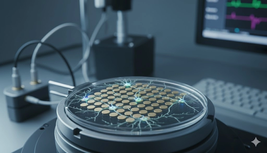
The Architecture of Multi-Electrode Arrays: Substrates and Sensors
Principles of Neural Network Electrophysiology using Multi-Electrode Arrays
High-Throughput Applications: Multi-Electrode Arrays in Drug Discovery and Disease Modeling
MEAs in Drug Discovery
MEAs in Disease Modeling
Advanced Data Analysis and Interpretation in Multi-Electrode Array Systems
Future Advancements in Multi-Electrode Array Technology
Frequently Asked Questions about Multi-Electrode Arrays
Mar 02, 2026
Product News
Thermo Fisher Launches Fluid Ease™ Pro ClipTip™ Electronic Pipette with High-Resolution Touchscreen and Advanced Workflow Controls
The system combines programmable control, intelligent power management, and a secure tip attachment mechanism to support consistent, reproducible liquid handling.

Feb 26, 2026
Product News
Waters Launches Microflow LC Columns with MaxPeak Premier Technology for Higher Sensitivity and Lower Resource Consumption
The product launch reflects continued investment in chromatographic chemistries that address real-world bottlenecks in method development and routine analysis

Feb 19, 2026
Technical Insight
Implementing Hydrogen Generators for Clean Lab Gas Supply: Safety and Benefits
Transitioning to on-site gas production streamlines analytical workflows, eliminates hazardous cylinder storage, and supports facility sustainability.

Feb 19, 2026
Technical Insight
Waste Minimization in Laboratory Settings: Best Practices for Chemical, Biological, and Electronic Waste
Effective waste minimization in laboratories improves safety and sustainability. Explore methods for managing chemical, biological, and electronic waste.
Feb 19, 2026
Technical Insight
Circular Economy in Laboratory Equipment: How to Adopt Repair, Reuse, and Refurbishment Strategies
Adopting a circular economy for laboratory equipment minimizes environmental impact while maximizing instrument lifespan and optimizing laboratory budgets.
Feb 12, 2026
Product News
$5M+ in GMP Biopharma Manufacturing & QC Equipment Heads to Online Auction
A Southern California GMP manufacturing and clinical-stage QC laboratory is liquidating new and late-model equipment and unused inventory in an online auction opening February 11, 2026

Feb 12, 2026
Product News
2026 SLAS: Inspiration Catalyzing Discovery
A LabX look at the annual conference and exhibition, a key forum for networking, collaboration, and advancement of new technologies
Feb 11, 2026
Product News
Angstrom Scientific Named U.S. Distributor for Radalytica’s RadalyX Robotic X-ray and CT System
Angstrom Scientific, known for representing microscopy and materials analysis technologies across North America, adds Radalytica’s robotic X-ray and CT platform to its portfolio of analytical and imaging solutions.

Feb 10, 2026
Technical Insight
Eco-Friendly Refrigeration: Evaluating Natural Refrigerants and Low-GWP Cooling Systems
This guide explains the operational and environmental benefits of adopting natural refrigerants and low-GWP cooling systems in laboratory settings

Feb 10, 2026
Technical Insight
The Ultimate Guide to Sustainability in Laboratory Equipment and Operations
A comprehensive analysis of sustainable laboratory practices, focusing on energy-efficient equipment, waste management, and digital workflows