Remapping Research and Discovery with Spatial Imaging

Written By: Damon Anderson, PhD
Dec 03, 2025
Spatial imaging has become a powerful enabling technology for modern scientific discovery
At its core, spatial imaging answers a fundamental question traditional molecular techniques cannot: Where are biological signals happening within intact tissues? By preserving spatial context, researchers gain insight not only into what molecules are present but how they interact within their native microenvironment—a critical factor in understanding health and disease.
Conventional bulk and single-cell methods average signals or dissociate cells. This averaging erases the architecture that underlies and drives biological function. Spatial imaging restores that architecture, enabling scientists to visualize cell types, molecular gradients, and structural features with subcellular precision. This level of resolution is transforming fields ranging from cancer biology—where tumor–immune boundaries dictate therapeutic response—to neuroscience, where mapping synaptic organization reveals circuit-level mechanisms underlying behavior and neurodegeneration.
Advances in multiplexed fluorescence, mass spectrometry–based imaging, and spatial transcriptomics now allow researchers to interrogate dozens to thousands of targets within a single tissue section. These data-rich images support more accurate biomarker discovery, better disease classification, and improved translational models. They also accelerate drug development by showing how candidates affect specific cell populations and pathways in situ.
Ultimately, spatial imaging empowers scientists to move beyond lists of molecules and toward a holistic understanding of biological systems. By integrating molecular identity with spatial relationships, researchers can build more predictive models, uncover hidden cellular interactions, and generate insights that would remain invisible using conventional methods alone.
Read on and learn more about the techniques and solutions that are helping to push the forefront of this new paradigm of investigation.
Mar 02, 2026
Product News
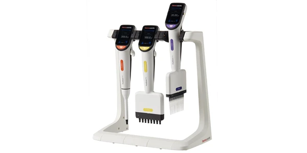
Thermo Fisher Launches Fluid Ease™ Pro ClipTip™ Electronic Pipette with High-Resolution Touchscreen and Advanced Workflow Controls
The system combines programmable control, intelligent power management, and a secure tip attachment mechanism to support consistent, reproducible liquid handling.
Feb 26, 2026
Product News
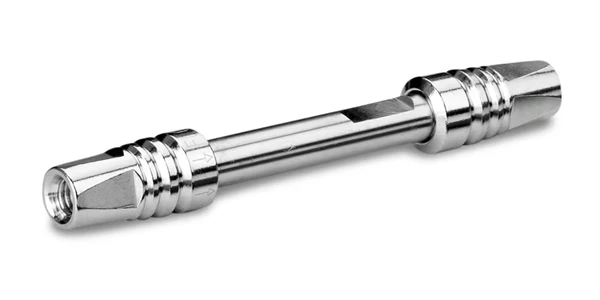
Waters Launches Microflow LC Columns with MaxPeak Premier Technology for Higher Sensitivity and Lower Resource Consumption
The product launch reflects continued investment in chromatographic chemistries that address real-world bottlenecks in method development and routine analysis

Feb 19, 2026
Technical Insight
Implementing Hydrogen Generators for Clean Lab Gas Supply: Safety and Benefits
Transitioning to on-site gas production streamlines analytical workflows, eliminates hazardous cylinder storage, and supports facility sustainability.
Feb 19, 2026
Technical Insight
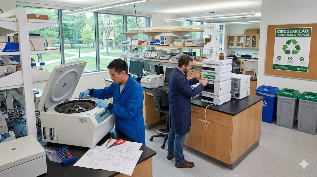
Circular Economy in Laboratory Equipment: How to Adopt Repair, Reuse, and Refurbishment Strategies
Adopting a circular economy for laboratory equipment minimizes environmental impact while maximizing instrument lifespan and optimizing laboratory budgets.
Feb 12, 2026
Product News
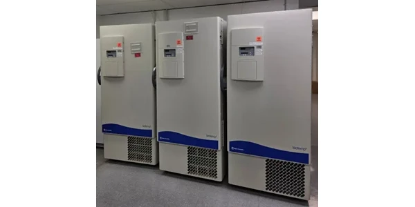
$5M+ in GMP Biopharma Manufacturing & QC Equipment Heads to Online Auction
A Southern California GMP manufacturing and clinical-stage QC laboratory is liquidating new and late-model equipment and unused inventory in an online auction opening February 11, 2026

Feb 12, 2026
Product News
2026 SLAS: Inspiration Catalyzing Discovery
A LabX look at the annual conference and exhibition, a key forum for networking, collaboration, and advancement of new technologies
Feb 11, 2026
Product News
Angstrom Scientific Named U.S. Distributor for Radalytica’s RadalyX Robotic X-ray and CT System
Angstrom Scientific, known for representing microscopy and materials analysis technologies across North America, adds Radalytica’s robotic X-ray and CT platform to its portfolio of analytical and imaging solutions.
Feb 10, 2026
Technical Insight
Eco-Friendly Refrigeration: Evaluating Natural Refrigerants and Low-GWP Cooling Systems
This guide explains the operational and environmental benefits of adopting natural refrigerants and low-GWP cooling systems in laboratory settings
Feb 10, 2026
Technical Insight
The Ultimate Guide to Sustainability in Laboratory Equipment and Operations
A comprehensive analysis of sustainable laboratory practices, focusing on energy-efficient equipment, waste management, and digital workflows
Jan 12, 2026
Technical Insight
The Best Polarimeters of 2026
A comprehensive guide to the top-rated polarimeters of 2026, featuring high-speed, high-precision, and budget-friendly models for every laboratory application.