Latest Innovations in Optogenetics and Electrophysiology Integration

Written By: Craig Bradley
Oct 15, 2025
GEMINI (2025) The precise manipulation and simultaneous monitoring of biological function are central requirements for modern neuroscientific and cardiac research. The coupling of optogenetics, which provides cellular specificity and millisecond-level temporal resolution in controlling genetically defined cell populations, with electrophysiology integration, which offers the gold standard for measuring real-time electrical activity, represents a major technical leap. This synergistic combination is crucial for moving beyond correlation to establish definitive causal relationships within complex neural circuits, and recent advancements in probe design, fabrication, and data analysis are rapidly expanding the scope and scale of achievable experiments in the modern laboratory setting. The scale and complexity of neural circuits demand recording techniques capable of capturing activity from hundreds of neurons simultaneously while applying localized light stimulation. Traditional methods relying on separate optical fibers and low-channel count electrodes often compromise spatial specificity, leading to activation of non-targeted cell types or volume illumination effects. A major innovation addressing this challenge is the development of next-generation high-density silicon probes that monolithically integrate light delivery structures with electrical recording sites, fundamentally improving electrophysiology integration. The introduction of specialized multimodal probes, such as the Neuropixels Opto, exemplifies this integration shift. These devices combine hundreds of electrical recording sites (up to 960) with multiple micro-LED (µLED) emitters onto a single, narrow shank. This arrangement allows for depth-specific and spatially addressable optogenetic stimulation, overcoming the limitations of broad-field illumination. Key advantages of these high-density integrated probes include: Cellular Specificity and Optotagging: By combining high-resolution spiking data with precise light delivery, researchers can definitively identify (optotag) specific, genetically modified neurons whose activity is being recorded. This is achieved by observing the recorded electrical response immediately following a brief light pulse. Probes with multiple light sets (e.g., blue and red) facilitate the parallel identification of two distinct cell populations expressing different light-sensitive opsins. Reduced Photothermal Artifacts: The highly localized nature of the light delivery, combined with the efficient design of the silicon substrates, minimizes unwanted thermal artifacts that can confound electrophysiological measurements, particularly during chronic in vivo recordings. Chronic and Freely Behaving Studies: Miniaturization and advanced packaging techniques now enable the use of these complex optoelectronic neural probes in freely moving animal models, offering unprecedented access to circuit function during naturalistic behavior. The resulting large-scale data sets provide a richer foundation for understanding dynamic circuit dynamics. Micro-LED (µLED) technology is transforming optogenetics by enabling light delivery with cellular-level spatial resolution and sub-millisecond temporal control. The integration of these micron-scale light sources directly onto the recording substrate represents a major step forward for fine-scale circuit dissection. µLED arrays embedded in silicon probes offer a distinct advantage over optical fibers by allowing independent control of light intensity and duration at multiple points along the recording shank. The distance between the light source and the targeted neuronal soma can be reduced to tens of micrometers, maximizing light efficiency and minimizing the effective volume of tissue stimulated. This close proximity allows for tunable illumination power, meaning the tissue volume activated by the opsin can be precisely matched to the tissue volume within the recording range of the adjacent microelectrodes. A significant technical hurdle in combined light and electrical experiments is spectral congestion and optical crosstalk, particularly when integrating optogenetics with optical sensing technologies. When genetically encoded indicators (GEIs) or voltage-sensitive fluorescent dyes (VSDs) are used alongside activating opsins (e.g., Channelrhodopsin-2, ChR2), the excitation/activation and emission spectra often overlap, resulting in contaminated readouts. Current solutions to mitigate this issue include: Strategic Opsins and Dyes Selection: Utilizing red-shifted excitatory opsins (e.g., Chrimson, ReaChR) or inhibitory opsins (e.g., GtACR) combined with blue-light-activated GEIs or VSDs to separate the spectral windows. Isosbestic Point Exploitation: Employing advanced techniques, such as using voltage-sensitive dyes and calcium indicators and operating near the isosbestic point of one dye, allows for simultaneous monitoring and manipulation with minimal crosstalk, yielding clean electrophysiology integration data. Advanced Micro-Optics: Developing sophisticated projection and micro-optics systems to improve the fill factor and optical efficiency of µLED arrays, ensuring the necessary irradiance reaches the target neurons without excessive power consumption or broad scattering. While high-density probes dominate in vivo innovation, in vitro and high-throughput cellular screening relies on innovative electrophysiology integration platforms, particularly those centered around Automated Patch Clamping (APC) and Microelectrode Arrays (MEAs). APC systems, traditionally used for high-throughput drug screening of ion channels, have been adapted to incorporate localized optogenetic stimulation. These platforms utilize planar patch-clamp chips integrated with light sources, typically high-intensity LEDs or light guides, allowing for rapid optical control of ion channels expressed in cell lines. This integrated approach offers crucial benefits in preclinical and cellular studies: Drug Target Validation: Optogenetically modified cell lines expressing specific channelrhodopsins can be simultaneously voltage-clamped and optically stimulated. This enables high-throughput screening of pharmacological compounds that modulate ion channel activity, providing precise mechanistic data that complement traditional electrical stimulation. All-Optical Approaches: The combination of optogenetic actuators and sensors (GEIs like GCaMP for calcium or GEVs for voltage) facilitates entirely contact-free methods for monitoring and manipulating cardiac or neuronal activity. These all-optical systems bypass the need for physical electrodes, reducing mechanical perturbation and enabling high-content screening. MEAs with Optical Stimulation: Standard MEA plates are increasingly paired with integrated optical systems for synchronized video, electrical recording, and light stimulation. Specialized MEA designs, such as perforated MEAs (pMEAs) and mesh MEAs, further enhance tissue-electrode coupling and nutrient exchange for long-term culture of organoids or slices, allowing for long-term, multimodal opto-electrophysiology studies. The combination of multi-site recording and precise spatiotemporal light perturbation generates vast, complex, and high-dimensional datasets. The success of optogenetics experiments is increasingly dependent on the algorithms and computational methodologies used to manage, process, and interpret this combined data. A persistent technical challenge is the light-induced electrical artifact that overlaps with the neuronal response. When the stimulating light is delivered, the photodiode effect, temperature changes, and changes in junction potential can generate a large electrical artifact that corrupts the recorded electrophysiological signal, especially in current-clamp mode. Advanced computational solutions are necessary for clean electrophysiology integration data extraction: Adaptive Subtraction Methods: Sophisticated algorithms use blank trials (lacking opsin expression) or pre-stimulus baseline recording to model and subtract the optical artifact, revealing the underlying physiological response. Principal Component Analysis (PCA) and Independent Component Analysis (ICA): These techniques are employed to separate the characteristic waveforms of the light artifact from true biological signals, significantly improving the signal-to-noise ratio during and immediately following stimulation. High-Pass Filtering: Utilizing hardware or software-based high-pass filters can effectively mitigate slow voltage drifts associated with thermal effects, though care must be taken to avoid distorting actual biological responses. The primary goal of integrating optogenetics with electrical recording is the causal dissection of neural circuits. Sophisticated analytical pipelines are now mandatory for inferring functional connectivity from the experimental data. Tools such as channelrhodopsin-assisted circuit mapping (CRACM) have long relied on patch-clamp recordings to precisely measure the strength and kinetics of synaptic connections. When applied to multi-site high-density data, specialized algorithms are required to: Identify Optogenetically Driven Spikes: Use statistical methods to verify that recorded spikes are causally linked to the optical stimulus and thus belong to the genetically targeted population. Model Synaptic Dynamics: Employ generalized linear models (GLMs) or dynamic causal modeling (DCM) to map the flow of information between perturbed and non-perturbed neuronal groups, quantifying the speed and sign (excitatory or inhibitory) of functional connections. Closed-Loop Systems: Integration requires real-time processing capabilities (often using Field-Programmable Gate Arrays, FPGAs) to detect specific electrical activity patterns and trigger an immediate optogenetic intervention, enabling the study of dynamic circuit function in a truly closed-loop, causal manner. The continuous evolution of integrated devices, from high-density in vivo probes to high-throughput in vitro APC systems, is providing the technical foundation necessary for comprehensive causal circuit analysis. The current trajectory in optogenetics and electrophysiology integration emphasizes higher spatial precision, greater channel count, and the reduction of experimental artifacts, moving the field toward truly large-scale, mechanistic studies of biological function. This technological convergence is not only accelerating the rate of discovery in basic neuroscience but is also providing critical tools for the preclinical screening of therapeutics targeting electrophysiological disorders. What is the core advantage of optogenetics combined with electrophysiology? How do Neuropixels probes advance electrophysiology integration?
Neuropixels Opto probes, and similar high-density silicon devices, integrate hundreds of electrical recording sites with micro-LED light emitters onto a single physical structure. This innovation allows for spatially addressable light delivery and large-scale, depth-specific recording, dramatically enhancing the fidelity of in vivo electrophysiology integration by minimizing light scattering and maximizing neuronal yield. What is spectral congestion in optogenetics research?
Spectral congestion refers to the overlap between the excitation/activation spectra of optogenetic actuators (light-sensitive proteins used for stimulation) and the excitation/emission spectra of optogenetic sensors (genetically encoded indicators or dyes used for monitoring). This overlap creates optical crosstalk, which can contaminate the electrophysiological or optical readout, necessitating careful selection of opsin-sensor pairs. Where is automated patch clamping (APC) used in integrated opto-electrophysiology?
APC systems, which enable high-throughput electrophysiological measurements in cell lines, are modified for integrated optogenetics by incorporating internal light sources. This setup is crucial in preclinical laboratories for fast, precise screening of ion channel function and drug compound effects, where light can activate specific opsins to induce or suppress channel activity. This article was created with the assistance of Generative AI and has undergone editorial review before publishing.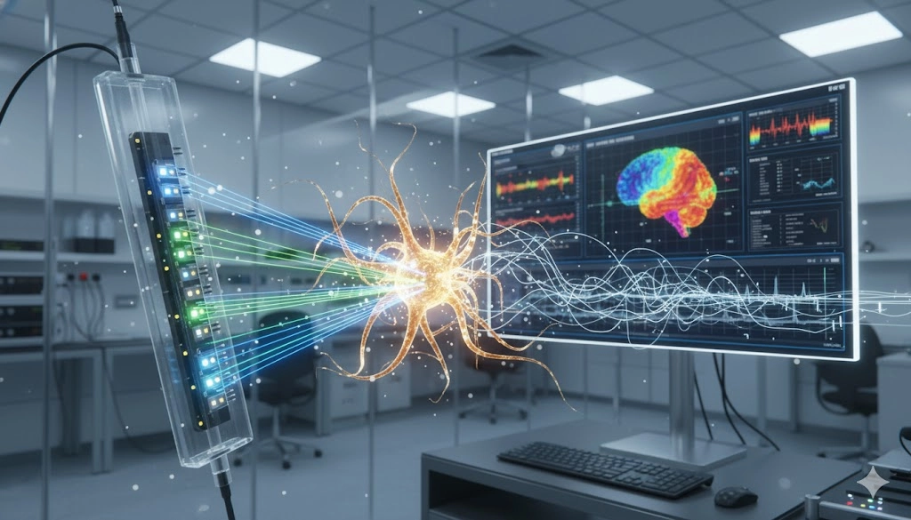
Advancing In Vivo Analysis with High-Density Opto-Electrophysiology Integration
Harnessing Micro-LED Array Systems for Precision Optogenetic Control
Overcoming Spectral Congestion in Opto-Electrophysiology
Integrated Platforms: Automated Patch Clamping and All-Optical Assays
Computational and Data Analysis Methodologies for Opto-Electrophysiology Data
Addressing the Stimulus Artifact
Causal Circuit Mapping and Connectivity Inference
Seamless Integration of Optogenetics and Electrophysiology for Mechanistic Discovery
Frequently Asked Questions on Optogenetics and Electrophysiology Integration
The primary advantage of integrating optogenetics with electrical recording is the ability to move beyond correlational studies to establish causal relationships in biological circuits. Optogenetics offers cell-type specificity and precise temporal control over activity, while electrophysiology provides a direct, high-fidelity readout of the resulting electrical changes.

Jan 09, 2026
Product News
BRANDTECH Scientific Introduces the Transferpette® pro Micropipette: A New Twist on Comfort and Control
The latest addition to its trusted line of precision liquid handling instruments, engineered for comfort, control, and repeatable accuracy.

Jan 06, 2026
Featured, Technical Insight
New Research Frontiers Unlocked by Next-Generation Flow Cytometry
Next-generation flow cytometry systems using acoustic focusing are opening entirely new areas of research that were previously impractical or inaccessible with conventional flow cytometers.

Jan 05, 2026
Buying Guides
The Bottom Line: How Smart Microcentrifuge Tube Choices Impact Lab Efficiency and Cost
Innovative microcentrifuge tube solutions are more than just lab consumables—they’re strategic tools that can reduce costs, improve outcomes, and support long-term sustainability goals.

Jan 05, 2026
Buying Guides
Future-Proofing Your Lab with Innovative Microcentrifuge Tube Solutions
As laboratories evolve to meet the growing demands of modern science, even the most basic tools—like microcentrifuge tubes—are being reimagined.

Jan 05, 2026
Buying Guides
Microcentrifuge Tube Innovations: Driving Sustainable Change
Integrating next-generation consumables into standard workflows can lead to a more sustainable and responsible research environment.

Jan 05, 2026
Buying Guides
Single Use Plastics: How to Manage these Major Contributors to Lab Waste
Reducing lab waste isn’t just a sustainability issue—it’s a financial and operational one, too.

Jan 05, 2026
Buying Guides
Common Microcentrifuge Tube Challenges in Sensitive Molecular Workflows
Key factors that can impact the performance of microcentrifuge tubes for PCR, qPCR, and NGS and other sensitive applications

Jan 05, 2026
Buying Guides
How to Research the Right Microcentrifuge Tubes for Your Application
Selecting the right microcentrifuge tubes starts with understanding the specific demands of your application.

Jan 05, 2026
Technical Insight
The Best Confocal Microscopes of 2026
Discover the best confocal microscopes of 2026 for advanced imaging. Compare top models from Zeiss, Evident, and Nikon for speed, resolution, and budget.

Dec 24, 2025
Featured, Popular Products, Technical Insight
Emerging Mass Spectrometry Technologies to Improve Intraoperative Diagnostics
Advances in mass spectrometry may soon reshape surgery, improving diagnostic accuracy and impacting post-operative outcomes