Comparing Flow Cytometry with Imaging Cytometry
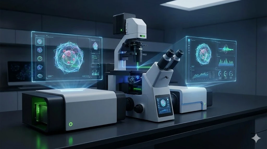
GEMINI (2025) In the modern laboratory, the analysis of individual cells is fundamental to understanding complex biological systems. Two powerful techniques, flow cytometry and imaging cytometry, are at the forefront of this effort. Although both methods provide data on cell populations, they operate on different principles and yield distinct types of information. Flow cytometry, with its origins in high-speed cell counting and sorting, is a time-tested technique for the quantitative analysis of thousands of cells per second. Imaging cytometry, a more recent evolution, combines the principles of flow cytometry with the power of fluorescence microscopy, capturing high-resolution images of each cell. The choice between these two methods is not a matter of one being superior, but rather of selecting the right tool for a specific research question. This guide explores the core principles, advantages, and limitations of each technique, providing a framework for making a strategic decision based on a laboratory’s analytical needs. Flow cytometry operates on the principle of hydrodynamically focusing a suspension of cells into a single-file stream. As each cell passes through one or more laser beams, it scatters light and emits fluorescence from conjugated probes. These signals are collected by an array of detectors, providing a rapid, high-throughput analysis. Quantitative Precision: The primary strength of flow cytometry lies in its ability to generate robust quantitative data on millions of cells in a short period. It precisely measures the intensity of fluorescent signals, allowing for accurate and reproducible quantification of protein expression, DNA content, and other cellular characteristics. This is particularly valuable for identifying distinct cell populations within a heterogeneous mixture and for tracking changes in marker expression over time or in response to a stimulus. High Throughput and Statistical Power: The speed of a flow cytometer is unparalleled. An instrument can analyze tens of thousands of cells per second, providing statistically powerful data from large sample populations. This high throughput makes flow cytometry ideal for large-scale screening applications, immunophenotyping of patient samples, and assessing the efficacy of drug candidates. The ability to quickly process large numbers of samples allows for the detection of rare cell populations with high confidence. Simplified Analysis: The data from a flow cytometer is typically presented as dot plots or histograms. While complex multi-color experiments require careful compensation and analysis, the data structure is relatively straightforward compared to the pixel-by-pixel information of images. This allows for rapid visualization of cell populations and a streamlined analysis workflow for routine assays. Imaging cytometry, also known as image-based cytometry, takes a fundamentally different approach. It captures a high-resolution image of each cell as it passes through the system. This technique preserves the spatial context and morphological information that are lost in traditional flow cytometry. Morphological and Subcellular Insight: A key benefit of imaging cytometry is its ability to provide detailed morphological information. Researchers can analyze a cell’s size, shape, and nuclear morphology, which can be critical for distinguishing between cell types or for assessing cellular health. Furthermore, imaging cytometry excels at determining the subcellular location of fluorescent markers. It can be used to quantify the translocation of transcription factors into the nucleus, the co-localization of proteins in a specific organelle, or the presence of a marker on the cell surface versus in the cytoplasm. Preservation of Spatial Relationships: Unlike flow cytometry, which disrupts the spatial arrangement of cells, imaging cytometry preserves the relationships between cells. This is particularly valuable for studying cell-cell interactions, such as T-cell and target-cell conjugates, or for analyzing the formation of clusters and rosettes. This capability provides a visual and spatial context that is impossible to obtain from single-cell flow data. Applications for Rare and Complex Events: Imaging cytometry is an excellent tool for analyzing rare or transient events. Because each cell is imaged, it is possible to manually review specific cells of interest. For example, in drug discovery, imaging cytometry can be used to identify a small population of cells undergoing a complex morphological change or to screen for cells that have successfully internalized a nanoparticle. Understanding the trade-offs between these two technologies is essential for optimizing a laboratory’s workflow. The table below summarizes the key differences in their operational characteristics and data output. Feature Flow Cytometry Imaging Cytometry Throughput High (10,000+ events/sec) Low to Medium (1-100 events/sec) Data Type Quantitative fluorescence intensity Quantitative fluorescence intensity, brightfield, morphology Information Gained Phenotype, cell count, protein expression level Phenotype, morphology, cell-cell interactions, subcellular localization Spatial Context Lost Preserved Best For High-throughput screening, cell counting, bulk phenotyping, sorting Rare event analysis, studies requiring morphological data, cell signaling, co-localization studies The decision between flow cytometry and imaging cytometry should be driven by the specific biological question. The two technologies are not in direct competition; instead, they are complementary tools that can be used to gain a more complete picture of cellular function. When to use Flow Cytometry: A laboratory should lean toward flow cytometry when the primary goal is to obtain statistically robust data on large populations. For example, an immunologist studying the prevalence of a specific T-cell subtype in a patient cohort would benefit from the speed and quantitative power of flow cytometry to analyze thousands of cells. When to use Imaging Cytometry: Imaging cytometry is the superior choice when the research question requires visual or spatial context. A researcher studying the activation of a transcription factor and its subsequent movement into the nucleus would need imaging cytometry to track this process at the single-cell level. Similarly, a lab investigating the morphological effects of a new drug would use imaging cytometry to analyze changes in cell size and shape. Often, the most powerful approach involves a hybrid workflow. Flow cytometry can be used as a high-throughput initial screen to identify a population of interest, which can then be sorted and analyzed in greater detail using imaging cytometry. This sequential approach allows for the benefits of both speed and spatial resolution. The choice between flow cytometry and imaging cytometry is a strategic decision that shapes a laboratory’s analytical capabilities. The two techniques, while distinct in their approach, are a powerful combination for cellular analysis. Flow cytometry provides the breadth of data needed for statistical confidence and large-scale studies, while imaging cytometry provides the depth and visual context necessary for understanding complex cellular processes. By recognizing the unique strengths of each method, laboratory professionals can select the right tool or, more effectively, integrate both technologies into their workflow to gain a more complete and accurate understanding of cellular biology. What is the main trade-off between the two technologies?
The primary trade-off is between throughput and information. Flow cytometry offers unparalleled speed and statistical power, while imaging cytometry provides rich morphological and spatial data at a lower throughput. Can imaging cytometry be used for cell sorting?
While some imaging cytometers can perform a form of sorting, it is typically not as fast or as precise as dedicated flow cytometers with sorting capabilities. Which technique is better for analyzing cells from a solid tissue?
Flow cytometry is the standard for analyzing cells from solid tissue, as it requires the tissue to be disaggregated into a single-cell suspension. Imaging cytometry can be used to analyze solid tissue sections, but this requires a different workflow and is typically done using traditional microscopy rather than a high-throughput imaging cytometer. Is imaging cytometry a replacement for a fluorescence microscope?
Imaging cytometry and fluorescence microscopy are complementary. While both provide image-based data, imaging cytometers are designed for high-throughput, automated analysis, whereas a microscope is often used for lower-throughput, more detailed manual inspection.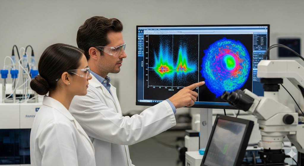
Flow Cytometry: The Speed and Scale Advantage
Imaging Cytometry: The Spatial and Morphological Advantage
A Comparative Overview of Cytometry Methods
Choosing the Right Tool for the Job
A Synergistic Approach in Cytometry
FAQ: Flow Cytometry vs. Imaging Cytometry
