Versatile and Efficient Benchtop Scanning Electron Microscopes

Written By: Damon Anderson, PhD
Nov 02, 2017
Downsizing has been a recent and growing trend in laboratory equipment. Not necessarily in the number or the scope of the instruments, rather, in the size of the instruments and the physical space in which they must operate. This is due to several reasons including the fact that laboratory space is becoming a precious commodity for cost reasons, forcing research staff to produce more and occupy less. In contrast, many labs are expanding both the number of instruments and the throughput of data acquisition, in part due to the nature of next generation applications such as deep sequencing and others. Furthermore, modern research labs encompass a greater scope of analytical techniques and specialty fields than ever before, adding yet another factor to the lab space equation.
Efficiency has emerged as a top priority for labs in considering, not just the space issue, but for equipment performance, ease of use and maintenance, and compatibility with data processing streams as well. When it comes to high complexity analytical instruments, there is always a balance between performance and efficiency. This is particularly applicable to the electron microscope, a technology that has traditionally been associated with large, complex, custom suited facilities – a concept that runs in direct contrast to this new age of downsized efficiency
The scanning electron microscope or SEM microscope has evolved to meet many of the requirements discussed above. The instrument and accessories have been effectively miniaturized to benchtop scale without sacrificing the features that make the technology powerful for so many research applications.
SEM was commercialized in the 1960's, and since then has assisted researchers in exploring novel frontiers in biology and material science, uncovering a miniature world previously unknown to optical microscopy. There was traditionally a divide between the two techniques, optical versus electron microscopy. Tissue or live cells were imaged by light microscopy, and then prepared through rigorous sample treatment methods prior to more laborious SEM imaging.
Introduction of the NeoScope technology from JEOL instruments supported a new concept that complemented both optical microscopes and traditional SEMs in the lab. The technology brought SEM to the benchtop while enabling seamless integration of optical microscope features into a higher resolution platform with up to 60,000X magnification. The same samples that are examined via light microscopy can be interrogated with much higher depth of field and resolution, importantly without additional extensive sample preparation. Many optical microscope features such as simplicity of use and size footprint are maintained while offering advanced settings for pre-screening of biological and composite samples and others – applications which were traditionally the domain of larger SEM instruments.
The new JEOL JCM-6000 NeoScope Versatile Benchtop SEM features a modern design with state-of-the-art features. Operation is via a touchscreen and is simplified with auto focus, auto alignment, auto contrast, and auto brightness controls. The instrument functions under both low and high vacuum modes with three settings for voltage acceleration. These features enable a variety of applications which can be programmed via pre-stored recipe files.
This new NeoScope offers both an Everhart Thornley type SE detector as well a high sensitivity solid state back scattering electron (BSE) detector. A full-featured Energy-dispersive X-ray Spectroscopy (EDS) with SDD technology is available for advanced analytical applications.
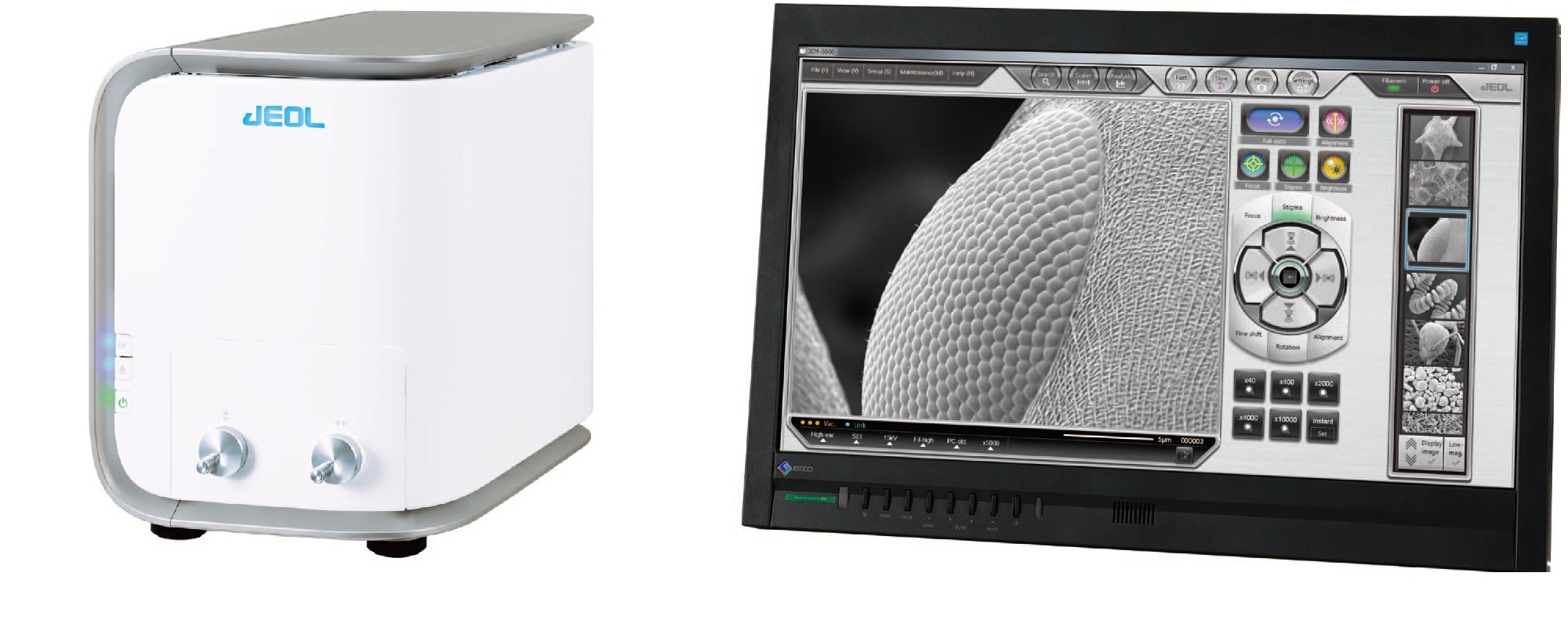
This NeoScope SEM microscope incorporates many important features and functions that make it both an efficient and powerful device. The touchscreen access and easy to use application software make it accessible by a variety of users and streamlined for processing of rich sample throughput and data streams. Tilting and rotating functions permit viewing at different angles, resulting in 3-D information on the sample. Many additional capabilities make this platform valuable for a variety of uses from neuroscience, to materials science, to manufacturing processes.
Whether used by trained electron microscopists as a simple screening instrument, or by lab technicians as a higher resolution alternative to the light microscope, the NeoScope accelerates the overall pace of research as is a model of imaging efficiency.
View JEOL NeoScope listings on LabX.com
The resolution of the electron microscope, scanning electron magnification, sem microscope uses, and specimen preparation for scanning electron microscopy are topics for other articles. Take a closer look at the LabX microscopy resources for more insight:
What is a Limitation of Using Electron Microscopes to View Specimens?
Brightfield Light Microscopy: Technical Insights and Applications
Fluorescence Light Microscopy: Technical Insights and Applications

Dec 24, 2025
Featured, Popular Products, Technical Insight
Emerging Mass Spectrometry Technologies to Improve Intraoperative Diagnostics
Advances in mass spectrometry may reshape surgery, improving diagnostic accuracy and impacting post-operative outcomes
Dec 23, 2025
Technical Insight
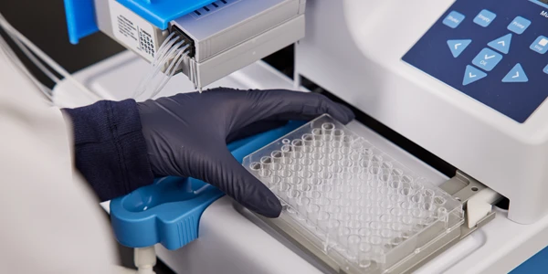
The Best Microplate Washers/Dispensers of 2026
Discover the top microplate washers/dispensers for 2026. Compare the latest models from Agilent, BlueCatBio, and Thermo Fisher to optimize your lab's throughput and precision today.
Dec 15, 2025
Featured, Popular Products
Automated 3D Cell Culture Workflows: Reshaping Disease Research and Drug Development
As 3D cell systems move from exploratory research into routine use across drug development and translational science, automation is becoming essential.
Dec 11, 2025
Technical Insight
The Best FPLC Systems of 2026
Discover the top FPLC systems of 2026 for protein purification. This guide reviews price, specs, and performance for leading lab equipment models.
Dec 11, 2025
Technical Insight
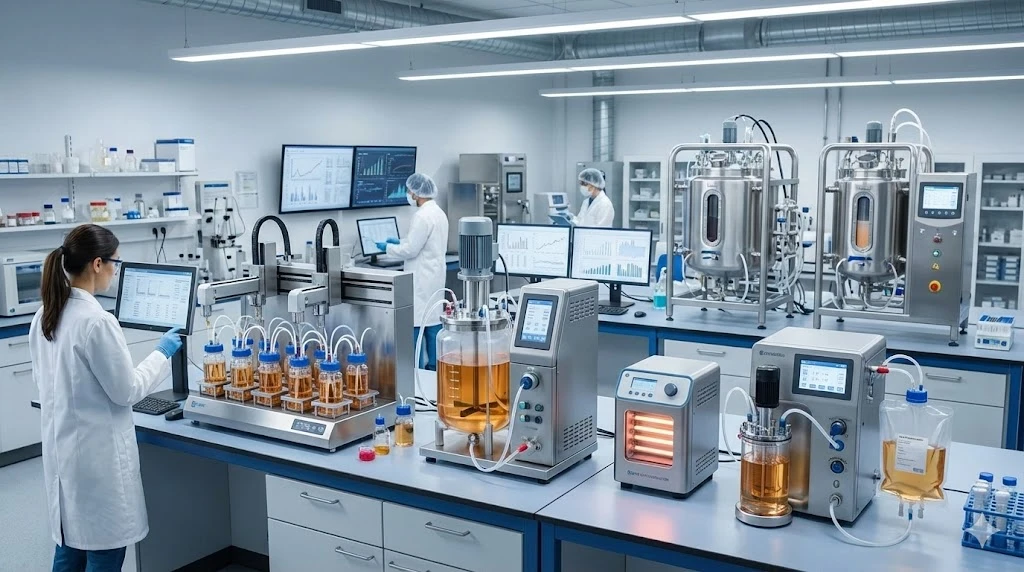
The Best Fermenters/Bioreactors of 2026
Explore the top fermenters/bioreactors of 2026 for research and production. Discover high-throughput, single-use, and budget-friendly models to optimize your lab's workflow.
Dec 11, 2025
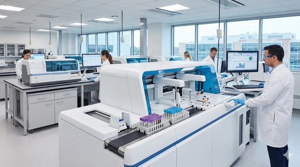
The Best ELISA Kits and Immunoassay Systems of 2026
Discover the top-rated ELISA kits and immunoassay systems of 2026. We review high-throughput analyzers and budget-friendly models for modern labs.
Dec 10, 2025
Featured, Popular Products
How Microplate Dispensers Enable Modern Research and Discovery
From drug discovery to genomics, almost every lab today depends on microplate dispensers.
Dec 08, 2025
Technical Insight
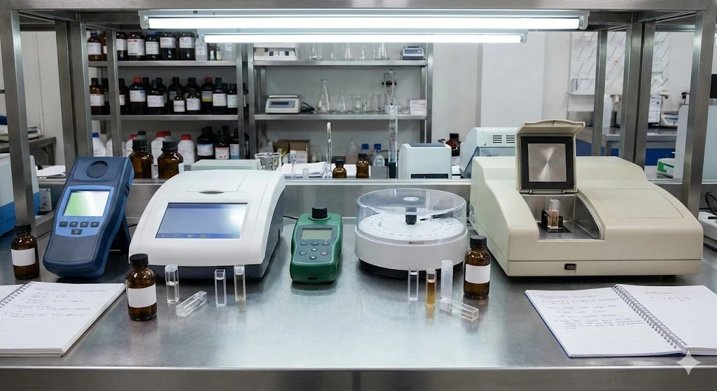
The Best Colorimeters of 2026
Discover the top-rated colorimeters of 2026 for laboratory professionals. We review the best new models for accuracy, speed, and chemical analysis.
Dec 08, 2025
Technical Insight
The Best DNA Sequencers of 2026
Discover the top-performing DNA sequencers of 2026. This guide reviews the latest high-throughput, benchtop, and long-read models for modern laboratories.
Dec 08, 2025
Technical Insight
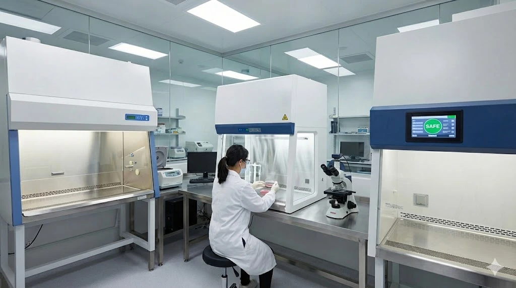
The Best Biosafety Cabinets and Laminar Flow Hoods of 2026
Discover the top biosafety cabinets and laminar flow hoods of 2026. This guide reviews safety, efficiency, and features for optimal laboratory protection.