A Modern Guide to Light Microscopy: Components, Techniques, and Innovations

Written By: Craig Bradley
May 04, 2017

ImageFX (2025)
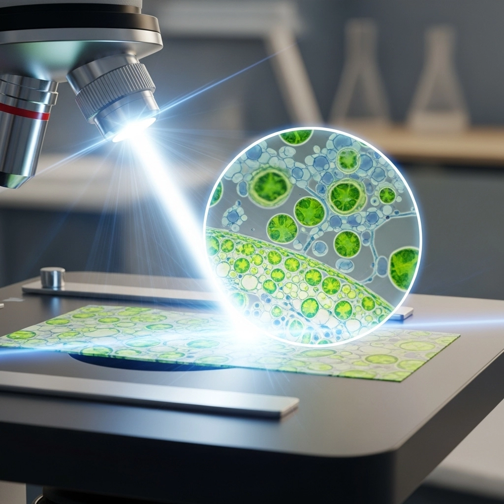
From the early observations of Antonie van Leeuwenhoek to today’s high-tech digital imaging systems, the field of light microscopy has undergone an extraordinary transformation. Modern microscopes not only offer incredible clarity and resolution but also unlock the potential to observe and analyze the tiniest aspects of biological and material sciences. Whether you're a seasoned researcher or a curious student, understanding the essentials and advancements in light microscopy is critical.
Key Components of a Light Microscope
Understanding the fundamental structure of a light microscope is crucial for proper usage and effective imaging. Modern light microscopes typically consist of the following elements:
1. Eyepiece (Ocular Lens)
Typically provides 5x to 20x magnification
Works in tandem with objective lenses to sharpen the image
2. Objective Lenses
Primary lenses that magnify the specimen
Characterized by:
Magnification Power (e.g., 10x, 40x, 100x)
Numerical Aperture (NA): Measures light-gathering ability and resolution
3. Focus Mechanism
Coarse and fine focus knobs adjust the stage to bring samples into clear view
4. Stage and Frame
Stage: Platform that holds the slide, includes an aperture for light
Frame: Rigid body that supports the entire setup
5. Illumination System
Traditional: Adjustable halogen lamps
Modern: LED or laser-based light sources
Compatible with multiple contrast-enhancing techniques like:
Cross-polarized light
Dark-field
Phase-contrast
Brightfield Microscopy: Classic Yet Powerful
Brightfield imaging remains one of the most widely used methods in optical microscopy. It's ideal for fixed, stained specimens and offers simplicity with powerful imaging capability.
Advantages of Brightfield Microscopy:
Works well with stained samples that absorb light
Delivers colored or dark images on a bright background
Can be adapted for viewing live specimens (though staining may be necessary)
Best Practices:
Use appropriate stains to highlight cellular structures
Adjust light intensity and diaphragm for optimal contrast
Explore our full guide to staining techniques to enhance your imaging workflow.
Advancements in Light Microscope Imaging
Brightfield may be traditional, but it now pairs seamlessly with modern imaging technologies for enhanced performance.
Digital Imaging Systems
Capture high-resolution images with advanced digital cameras
Easily document and share observations for collaboration
3D Imaging Accessories
Enable real-time 3D visualizations of samples
Provide depth and spatial orientation not available in 2D views
Video Imaging
Records live interactions of motile organisms
Perfect for behavioral and environmental studies
Brightfield Meets Software
Modern software tools can:
Analyze cells without requiring fluorescence
Identify nuclei and cell boundaries using multi-focal plane images
Benefits of Software Integration:
Reduces need for fluorescence imaging
Minimizes color distortion from overlapping fluorescent channels
Clinical and Diagnostic Applications
Light microscopy remains essential in pathology and clinical diagnostics. The ability to examine and compare tissue samples provides critical insights into disease progression.
Real-World Application:
| Task | Role of Light Microscopy |
|---|---|
| Disease Diagnosis | Identifying abnormal tissue markers |
| Histopathology | Comparing stained tissues |
| Automated Analysis | High-throughput diagnostic workflows |
Modern laboratories now use automated microscopes capable of scanning and analyzing thousands of slides daily, improving accuracy and efficiency in diagnostics.
Final Thoughts on Light Microscopy
From its simple beginnings to its role in high-resolution imaging and diagnostics, light microscopy continues to be a cornerstone of modern science. As new technologies emerge, this essential tool becomes even more powerful, versatile, and accessible.
Explore top-tier microscopy systems, imaging tools, and diagnostic instruments at LabX.
FAQ on Light Microscopy
What are the main components of a light microscope?
Eyepiece, objective lenses, focus knobs, stage, frame, and illumination system form the essential parts of a light microscope.
How does brightfield microscopy work?
It transmits light through a sample, which absorbs or scatters it, producing a visible image on a bright background.
Can you use light microscopy for live cells?
Yes, but often requires staining or contrast techniques like phase-contrast or dark-field to improve visibility.
What is the role of numerical aperture in microscopy?
It determines the resolving power and light-gathering ability of objective lenses, impacting image clarity.

Jan 09, 2026
Product News
BRANDTECH Scientific Introduces the Transferpette® pro Micropipette: A New Twist on Comfort and Control
The latest addition to its trusted line of precision liquid handling instruments, engineered for comfort, control, and repeatable accuracy.

Jan 06, 2026
Featured, Technical Insight
New Research Frontiers Unlocked by Next-Generation Flow Cytometry
Next-generation flow cytometry systems using acoustic focusing are opening entirely new areas of research that were previously impractical or inaccessible with conventional flow cytometers.

Jan 05, 2026
Buying Guides
The Bottom Line: How Smart Microcentrifuge Tube Choices Impact Lab Efficiency and Cost
Innovative microcentrifuge tube solutions are more than just lab consumables—they’re strategic tools that can reduce costs, improve outcomes, and support long-term sustainability goals.

Jan 05, 2026
Buying Guides
Future-Proofing Your Lab with Innovative Microcentrifuge Tube Solutions
As laboratories evolve to meet the growing demands of modern science, even the most basic tools—like microcentrifuge tubes—are being reimagined.

Jan 05, 2026
Buying Guides
Microcentrifuge Tube Innovations: Driving Sustainable Change
Integrating next-generation consumables into standard workflows can lead to a more sustainable and responsible research environment.

Jan 05, 2026
Buying Guides
Single Use Plastics: How to Manage these Major Contributors to Lab Waste
Reducing lab waste isn’t just a sustainability issue—it’s a financial and operational one, too.

Jan 05, 2026
Buying Guides
Common Microcentrifuge Tube Challenges in Sensitive Molecular Workflows
Key factors that can impact the performance of microcentrifuge tubes for PCR, qPCR, and NGS and other sensitive applications

Jan 05, 2026
Buying Guides
How to Research the Right Microcentrifuge Tubes for Your Application
Selecting the right microcentrifuge tubes starts with understanding the specific demands of your application.

Jan 05, 2026
Technical Insight
The Best Confocal Microscopes of 2026
Discover the best confocal microscopes of 2026 for advanced imaging. Compare top models from Zeiss, Evident, and Nikon for speed, resolution, and budget.

Dec 24, 2025
Featured, Popular Products, Technical Insight
Emerging Mass Spectrometry Technologies to Improve Intraoperative Diagnostics
Advances in mass spectrometry may soon reshape surgery, improving diagnostic accuracy and impacting post-operative outcomes