

ImageFX (2025)
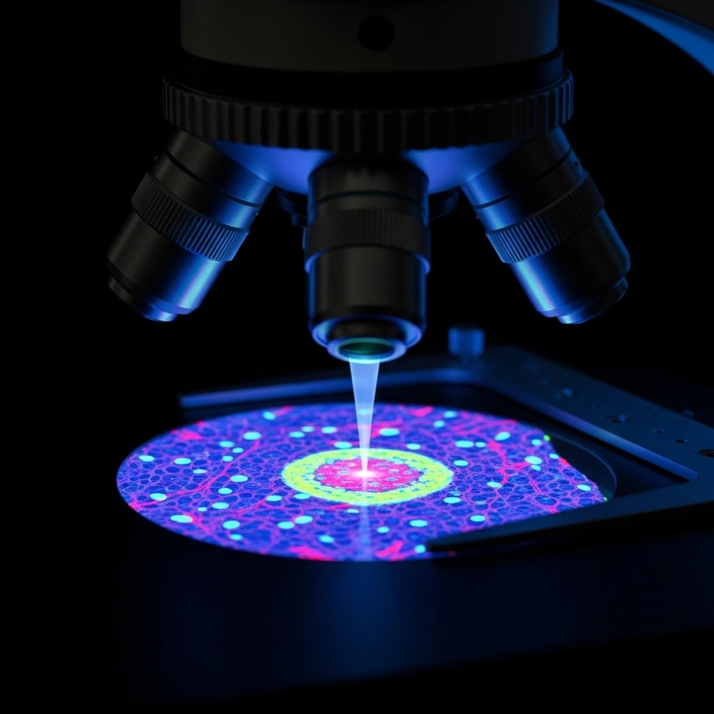
In the vast landscape of scientific exploration, the ability to visualize the intricate details of the microscopic world is paramount. While traditional optical microscopy provides foundational insights, fluorescence microscopy emerges as a powerful evolution, pushing the boundaries of what we can observe. Far more than just a light magnifier, this advanced technique harnesses the unique properties of fluorescent molecules to illuminate specific structures and processes within a sample, revealing a hidden universe of biological and material phenomena.
This guide delves into the core principles, diverse applications, and cutting-edge techniques that make fluorescence microscopy an indispensable tool across various scientific disciplines. From unraveling the mysteries of living cells to inspecting industrial materials, understand how this technology is transforming discovery.
How Fluorescence Microscopy Works: Key Principles and Components
At its heart, fluorescence microscopy operates on a fundamental principle: the interaction of light with specialized molecules called fluorophores. Unlike traditional brightfield microscopy, which relies on light absorption and scattering, fluorescence microscopy uses the phenomenon of fluorescence or phosphorescence for illumination.
Here's how it works:
Excitation: A sample containing fluorophores is illuminated with light of a specific, shorter wavelength (higher energy). This "excitation light" is precisely matched to the absorption spectrum of the fluorophores present.
Emission: Upon absorbing this energy, the fluorophores become excited. Almost instantaneously, they release this absorbed energy by emitting light at a longer, lower-energy wavelength. This emitted light is the "fluorescent signal" we observe.
Separation: A critical component of the microscope is a spectral emissions filter. This filter ensures that only the longer-wavelength emitted light from the fluorophores reaches the detector, effectively blocking the brighter, shorter-wavelength excitation light. This separation is crucial for achieving high contrast and clear images.
Common light sources for excitation include traditional xenon arc lamps and mercury-vapor lamps, though modern systems increasingly utilize highly efficient and precise LEDs and lasers, offering greater control and spectral purity. This innovative approach allows scientists to visualize structures and processes with remarkable clarity, often far beyond the resolution limits of conventional light microscopy.
Fluorophores and Advanced Fluorescence Imaging Techniques
The true magic of fluorescence microscopy lies in its ability to selectively highlight specific components within a complex sample. This is largely thanks to fluorophores – molecules that exhibit fluorescence. While some substances naturally auto-fluoresce, the scientific community widely employs a vast array of commercially available fluorophores. These engineered molecules boast well-defined excitation and emission spectra, allowing researchers to 'stain' or label specific structures, proteins, or molecules within a specimen.
The strategic selection of fluorophores is vital. By choosing fluorophores with distinct emission spectra, scientists can simultaneously identify and visualize multiple targets within a single sample, provided their emissions can be cleanly separated and distinguished from any background auto-fluorescence. This multiplexing capability is a cornerstone of modern biological research.
Building upon the basic fluorescence principle, several advanced microscopy techniques have emerged, each offering unique advantages for specific research questions:
Epifluorescence Microscopy: This is a widely used configuration, especially in life sciences. Both the illuminating light and the emitted fluorescent light pass through the same objective lens. This arrangement is particularly effective because reflected excitation light is efficiently filtered out by the objective, leading to a high signal-to-noise ratio and clear images.
Confocal Fluorescence Microscopy: A significant advancement, confocal microscopy uses a pinhole to block out-of-focus light, resulting in sharper, higher-resolution images, especially in thick specimens. It's an essential tool for studying 3D structures and dynamic processes in living cells.
Total Internal Reflectance Fluorescence (TIRF) Microscopy: TIRF provides extremely high signal-to-noise by illuminating only a very thin section of the sample near the coverslip. This is ideal for studying cell surface events and single-molecule dynamics.
Other Advanced Techniques: Fluorescence also enables sophisticated imaging methods like Förster Resonance Energy Transfer (FRET), Fluorescence Lifetime Imaging Microscopy (FLIM), Fluorescence Loss In Photobleaching (FLIP), and Fluorescence Recovery After Photobleaching (FRAP). These techniques provide quantitative information about molecular interactions, diffusion, and localization within living systems.
Applications of Fluorescence Microscopy in Research and Industry
The versatility of fluorescence microscopy makes it an indispensable tool across a broad spectrum of scientific and industrial fields. Its ability to visualize molecules and structures beyond the resolution limit of traditional light microscopes has revolutionized numerous areas:
Biological and Biomedical Sciences:
Immunology: Identifying and localizing immune cells and antigens.
Pathology: Diagnosing diseases by visualizing specific biomarkers in tissue samples.
Microbiology: Studying the morphology, dynamics, and interactions of bacteria, viruses, and other microorganisms.
Genetics: Mapping gene expression, chromosome structure, and DNA replication.
Cell Biology: Observing structural and molecular dynamics in living cells, including protein trafficking, organelle movement, and cellular signaling pathways.
Materials Science and Industrial Applications:
Geology: Identifying fluorescent minerals and understanding their composition.
Semiconductor Inspection: Detecting contaminants and impurities in microelectronic components.
Environmental Protection: Analyzing pollutants and their distribution in environmental samples.
The insights gained from fluorescence microscopy are critical for both fundamental research and practical applications, driving innovation in medicine, biotechnology, and beyond.
Enhancing Fluorescence Images with Deconvolution Software
Even with advanced microscopy techniques, images can sometimes contain "out-of-focus" information or scattered light that obscures fine details. This is where deconvolution plays a crucial role in enhancing image quality.
Deconvolution is a computational image processing technique that helps to exclude out-of-focus information from widefield microscope images. A widefield microscope, by its nature, detects light not only from the precise focal plane but also scattered light originating from other regions of the sample.
How Deconvolution Works: A sophisticated deconvolution software algorithm analyzes the raw image data and mathematically redirects fluorescent signals to their true origin. It essentially "undoes" the blurring caused by the microscope's optics and out-of-focus light.
Benefits: Compared to an original widefield image, a deconvoluted image reveals significantly sharper details, improves contrast, and allows for more precise localization of fluorescent signals. Crucially, it achieves this while preserving the quantitative data, enabling more accurate measurements and analyses. For instance, in the immunofluorescent imaging of HeLa cells (as shown in the source material), deconvolution allowed for a more precise determination of NF-κB molecule positions, highlighting its power in detailed cellular analysis.
Deconvolution is an essential post-acquisition processing step that transforms raw data into publication-quality images, maximizing the information extracted from your fluorescence microscopy experiments.
The Future of Fluorescence Microscopy: Innovations and Outlook
Fluorescence microscopy stands as a cornerstone of modern scientific research, offering unparalleled capabilities for visualizing the unseen. From its fundamental principles of light interaction and fluorophore application to its diverse array of advanced techniques and critical role in image optimization through deconvolution, this technology continues to drive breakthroughs across biological, medical, and material sciences. By providing a window into the molecular world, fluorescence microscopy empowers researchers to unravel complex processes, diagnose diseases, and innovate new solutions.
Ready to explore the tools that make these discoveries possible? Explore our full range of fluorescence microscopy equipment and accessories at LabX.
Frequently Asked Questions (FAQ)
What is the main advantage of fluorescence microscopy over traditional light microscopy? The primary advantage is its ability to visualize specific structures or molecules within a sample by labeling them with fluorophores. This provides high contrast and specificity, allowing researchers to observe phenomena that would be invisible or indistinguishable with traditional light microscopy.
How do fluorophores contribute to fluorescence imaging? Fluorophores are special molecules that absorb light at one wavelength and then emit it at a longer wavelength. They are used to selectively "stain" or tag specific components (like proteins or organelles) within a sample, making those components visible under the microscope when excited by the appropriate light source.
What are some key applications of fluorescence microscopy? Fluorescence microscopy is widely used in biological and biomedical sciences (e.g., immunology, pathology, cell biology, genetics), as well as in materials science, geology, and semiconductor inspection, for tasks like disease diagnosis, studying molecular dynamics, and detecting impurities.
Why is deconvolution important in fluorescence microscopy? Deconvolution is a computational technique that removes out-of-focus light and blurring from fluorescence images. It significantly enhances image clarity, sharpness, and signal-to-noise ratio, allowing for more precise localization of fluorescent signals and more accurate quantitative analysis of the data.


