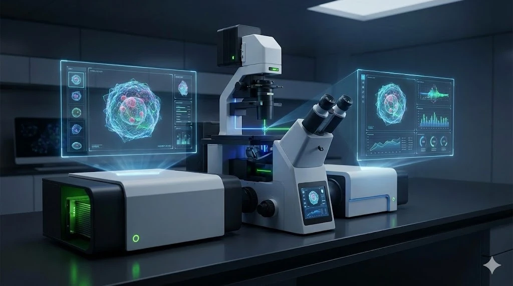
Capillary Electrophoresis: Principles and Applications

GEMINI (2025)
The demand for high-resolution analytical methods capable of characterizing complex biological and synthetic molecules has made capillary electrophoresis (CE) an indispensable technique in modern laboratory science. CE offers exceptional efficiency, speed, and minimal sample consumption, making it highly valuable for quality control, process monitoring, and research in the pharmaceutical and biotechnology industries.
This technique separates analytes based on their charge-to-mass ratio, using narrow-bore capillaries and high voltage to achieve ultra-high separation efficiencies that often exceed those of traditional chromatographic methods. Mastery of CE principles enables laboratory professionals to enhance workflow productivity and achieve rigorous analytical outcomes in both regulated and discovery environments.
Fundamental Principles of Capillary Electrophoresis
Capillary electrophoresis operates through two primary mechanisms: electrophoretic mobility and electroosmotic flow (EOF). Understanding how these forces interact is key to optimizing separation performance.
Electrophoretic Mobility (μₑₚ)
Electrophoresis describes the movement of charged particles in an electric field. The velocity of an ion (vₑₚ) is proportional to the field strength (E) and the ion’s electrophoretic mobility (μₑₚ):
vₑₚ = μₑₚ × E
Electrophoretic mobility depends on the analyte’s charge (q) and the frictional drag (f) it experiences in the buffer:
μₑₚ = q / f
-
Charge (q): Highly charged ions migrate faster than weakly charged ones. Buffer pH determines ionization state and thus effective charge—crucial for amphoteric analytes like peptides and proteins.
-
Frictional drag (f): Resistance encountered as ions move through the buffer, proportional to ion size and buffer viscosity. Smaller, more highly charged ions exhibit greater mobility.
Electroosmotic Flow (EOF)
EOF is the bulk movement of buffer solution through the capillary. The fused-silica inner wall contains silanol groups (–SiOH) that deprotonate at pH values above approximately 3, producing negatively charged surfaces (–SiO⁻). These attract positive counter-ions from the buffer, forming an electrical double layer (EDL).
When voltage is applied, the cations in the EDL migrate toward the cathode, dragging the entire buffer along. The EOF’s magnitude and direction determine how both charged and neutral species migrate.
-
Standard EOF (High pH): Typically directed toward the cathode, allowing all species—cations, anions, and neutrals—to reach the detector.
-
EOF Modulation: Adjusting the capillary wall (via coatings) or buffer composition allows control over EOF, enabling better separations.
The net analyte velocity (vₙₑₜ) combines electrophoretic and electroosmotic contributions:
vₙₑₜ = vₑₚ + vₑₒf
Neutral compounds migrate solely at EOF velocity, so their separation depends on extended methods such as Micellar Electrokinetic Chromatography (MEKC).
Capillary Electrophoresis Instrumentation and Method Development
A CE system is compact yet precise, comprising five core components:
-
Capillary: Fused-silica tubing (20–100 μm ID) ensures rapid heat dissipation, minimizing band broadening.
-
Buffer Reservoirs: Contain the running buffer, completing the electrical circuit.
-
High-Voltage Power Supply: Provides 10–30 kV, driving electrophoresis and EOF.
-
Injection System: Introduces nanoliter volumes of sample, either hydrodynamically (pressure) or electrokinetically (voltage).
-
Detector: Typically UV/Vis absorbance; alternatives include Laser-Induced Fluorescence (LIF) and Mass Spectrometry (MS).
Strategic Method Development
Method development in CE focuses on optimizing buffer chemistry, voltage, and capillary conditions to achieve the desired separation.
Key parameters include:
-
Running Buffer: Type, concentration, and pH influence both ionization and EOF strength.
-
Capillary Surface: Uncoated capillaries may cause adsorption issues; coatings such as polyacrylamide or PEG reduce EOF and improve reproducibility.
-
Temperature: Controlled between 15–40 °C to maintain consistent viscosity and EOF.
-
Voltage: Higher voltage accelerates separations but can increase Joule heating; balance is essential.
A structured approach typically begins with buffer screening, followed by optimization of ionic strength, voltage, and temperature, and finally, additives for selectivity or stability enhancement.
Capillary Electrophoresis vs HPLC
| Feature | Capillary Electrophoresis (CE) | High-Performance Liquid Chromatography (HPLC) |
|---|---|---|
| Separation Principle | Charge-to-mass ratio and size | Differential partitioning between mobile and stationary phases |
| Driving Force | Electric field | Hydraulic pressure |
| Efficiency | 100,000–1,000,000 plates | 10,000–100,000 plates |
| Band Broadening | Minimal | Significant (eddy diffusion, mass transfer) |
| Best Suited For | Charged molecules (proteins, peptides, ions, nucleic acids) | Neutral or non-polar small molecules |
| Mobile Phase | Simple aqueous buffers | Complex solvent mixtures |
Operational Advantages of CE
-
High Efficiency: EOF produces plug-like flow, minimizing dispersion and maximizing resolution.
-
Low Sample Consumption: Nanoliter volumes make CE ideal for scarce or costly samples.
-
Reduced Solvent Use: Minimal buffer volumes lower costs and environmental impact.
-
Orthogonality: CE provides a complementary mechanism to HPLC, enabling analysis of mixtures that resist chromatographic separation.
While HPLC offers greater load capacity and robustness for preparative work, CE excels in analytical-scale, high-resolution applications, especially for charged biomolecules.
Applications in Biotechnology, Pharmaceuticals, and Forensics
Pharmaceutical and Biologic Characterization
-
Purity and Impurity Profiling: CE ensures API quality through impurity analysis.
-
Chiral Separations: Cyclodextrin-based CE methods efficiently resolve drug enantiomers.
-
Biologics: CE techniques such as cIEF (for charge variants) and CGE (for size variants) are critical in monoclonal antibody characterization.
CE-MS in Proteomics and Metabolomics
Coupling CE with MS enhances detection sensitivity and resolution for complex samples.
-
Proteomics: CE-MS improves peptide mapping, PTM identification, and low-abundance protein detection.
-
Metabolomics: CE-MS effectively profiles highly polar metabolites poorly retained by HPLC.
Forensic Applications
Capillary electrophoresis is the gold standard for DNA and STR profiling.
- DNA Fragment Analysis: Polymer-filled capillaries separate STR fragments by size with high precision
- Automation: Enables high-throughput, quantitative forensic workflows
Selecting a Capillary Electrophoresis System
Choosing the right CE system involves aligning instrument features with analytical goals:
- Detection options: UV, LIF, or CE-MS interfaces depending on analyte type.
- Automation and Throughput: Automated rinsing, sampling, and multi-capillary systems enhance productivity
- Temperature Control: Critical for reproducibility and minimizing Joule heating.
- Compliance: Ensure software meets regulatory standards (e.g., 21 CFR Part 11)
Advanced Integration: CE-MS Interface
Sheath-liquid and sheath-gas interfaces stabilize electrospray ionization, merging CE’s separation power with MS’s identification capability. These hybrid systems are rapidly advancing in proteomics and metabolomics research.
Microchip Electrophoresis (ME)
Miniaturized systems integrate channels and detectors on a single chip, offering faster analysis, reduced sample needs, and portability—paving the way for point-of-care diagnostics.
Capillary Electrophoresis in the Modern Lab
Capillary electrophoresis remains a cornerstone of modern analytical science. Its unmatched efficiency, low sample and solvent requirements, and adaptability make it indispensable in biotechnology, pharmaceuticals, and forensics.
By mastering the fundamentals of electrophoretic mobility and EOF—and making informed choices in method development and system configuration—laboratories can leverage CE to meet the demanding standards of today’s analytical workflows. As CE technology evolves toward microchip and MS-integrated systems, its role in next-generation analytical platforms will continue to expand.
Frequently Asked Questions (FAQ)
What are the main challenges in CE method development?
Reproducibility can be affected by capillary wall interactions, EOF variability, and analyte adsorption. Stabilizing EOF through surface conditioning and buffer additives ensures consistent performance.
How does CE compare to HPLC for proteins and peptides?
CE offers superior resolution for charge variants and can preserve native protein structures under non-denaturing conditions, whereas HPLC provides higher loading capacity for preparative work.
How is CE used in forensic analysis?
CE separates fluorescently labeled STR fragments with high precision, forming the foundation of modern DNA profiling in human identification and paternity testing.
How does CE-MS enhance proteomics?
CE-MS combines CE’s high-efficiency separation of charged peptides with MS detection sensitivity, improving sequence coverage and post-translational modification analysis.
This article was developed with AI assistance and reviewed for technical and editorial accuracy before publication.
