Advancements in Mass Spec Imaging Presented at ASMS

Written By: Damon Anderson, PhD
Jul 06, 2022
Low micron-scale resolution and protein compound co-localization were two MALDI Imaging innovations showcased at the ASMS conference
The age of molecular-level imaging is upon us. What once was relegated to biophysical and microscopy techniques is now a domain shared by emergent imaging technologies. Not long ago, molecular imaging approaches using mass spectrometry were a more of a vision than reality.
The rapidly maturing field of MS Imaging is now experiencing increases in resolution and scope far beyond previous limitations. Several new solutions showcased at the American Society for Mass Spectrometry (ASMS) conference this year exemplify how far the field has come and where it is headed on the molecular imaging map.
MicroGRID timsTOF fleX
Bruker is a leader in MS Imaging technologies and incremental advancements in technology have brought MALDI Imaging to the forefront of innovation. In MALDI Imaging, spectra are collected at each point on a sample, which are then projected to create a 2-D spatial map. These maps can contain hundreds or thousands of unique spectral data points that can be used in discovery or characterization of the molecular and spatial details of a sample.
Advances in technology have enabled larger sample sizes, automated processing, and increases in feature resolution. This in turn has allowed imaging of tissue samples in rapid succession, disclosing details relevant to lipid profiling and small molecule distribution among other applications.
At ASMS this year, Bruker unveiled the next
iteration of MALDI Imaging instrumentation with the MicroGRID timsTOF fleX and
associated solutions.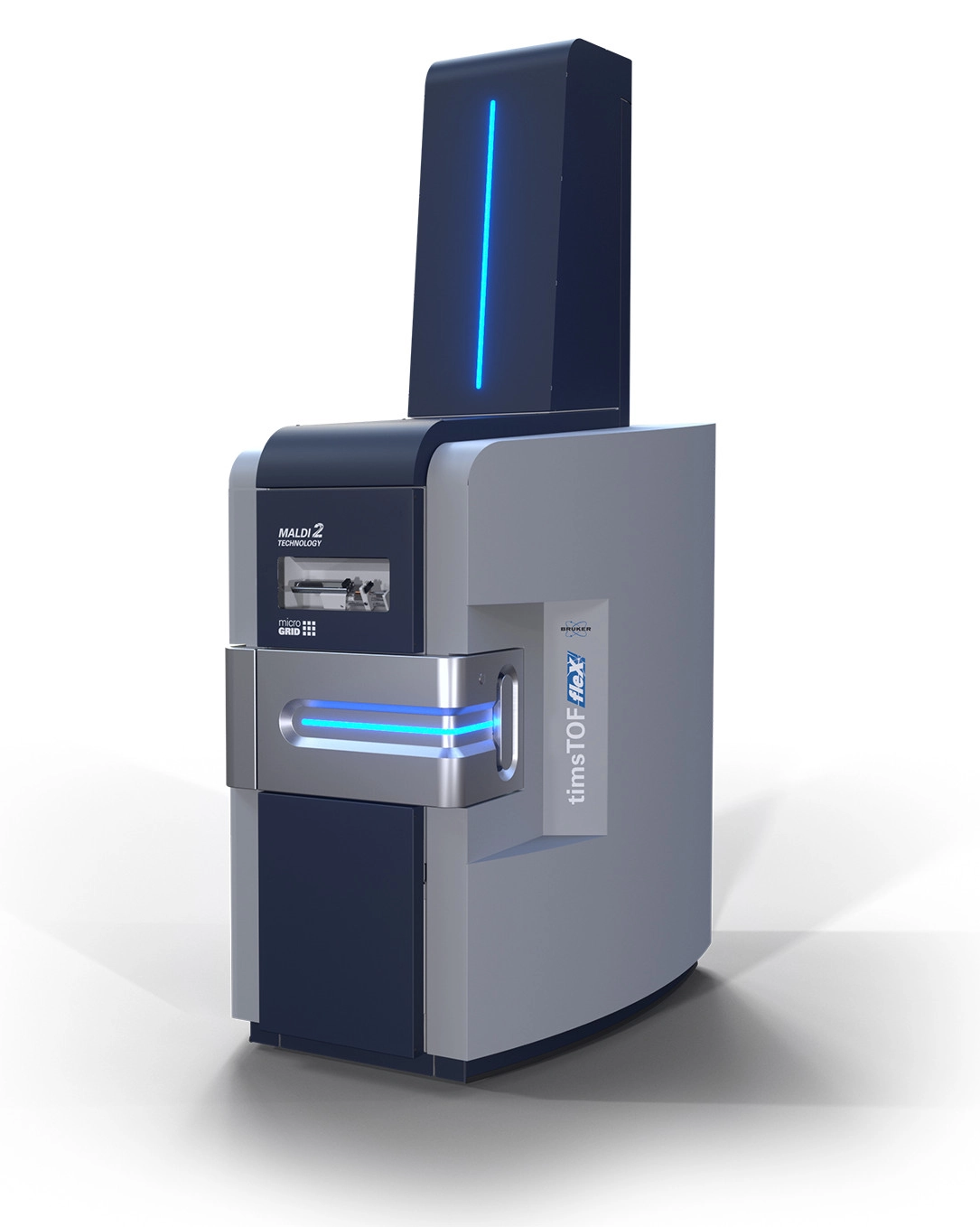
The new microgrid technology available on the timsTOF fleX permits spatial mapping down to 5-micron resolution.
- Laser position correction of microGRID offers improvements beyond mechanical limitations of Bruker’s previously released smartbeam 3D technology.
- The earlier SCiLS™ imaging software is upgraded on the latest platform to SCiLS™ autopilot, to allow out-of-the-box imaging without the need for adjustments to the setup.
- The new platform is also compatible with Bruker’s MALDI 2 ionization technology, which enhances the yield of sample ionization through a two-step laser desorption method. This feature frees up molecules typically confirmed to ion suppression and can produce up to 2-3 orders of magnitude greater sensitivity.
The potential applications for low micron mapping are many-fold. Subcellular features such as those implicated in disease versus healthy cells can be explored. Tissue and cellular penetration of drugs and metabolites can be imaged with higher resolution than previously. Cellular signaling pathways may be better defined using this new platform and suite of MALDI Imaging solutions.
MALDI HiPLEX-IHC
Keeping with a focus on mass spectrometry imaging innovations, Bruker announced the new MALDI HiPLEX-IHC platform at ASMS. This technology enables high-resolution imaging of specific protein types, among the backdrop of mass spec imaging of tissue sections.
Previous product development has enabled the use of high-resolution MALDI Imaging as a platform for analyzing fixed formalin paraffin embedded (FFPE) tissue sections. Bruker’s new strategic partnership with AmberGen builds upon this approach.
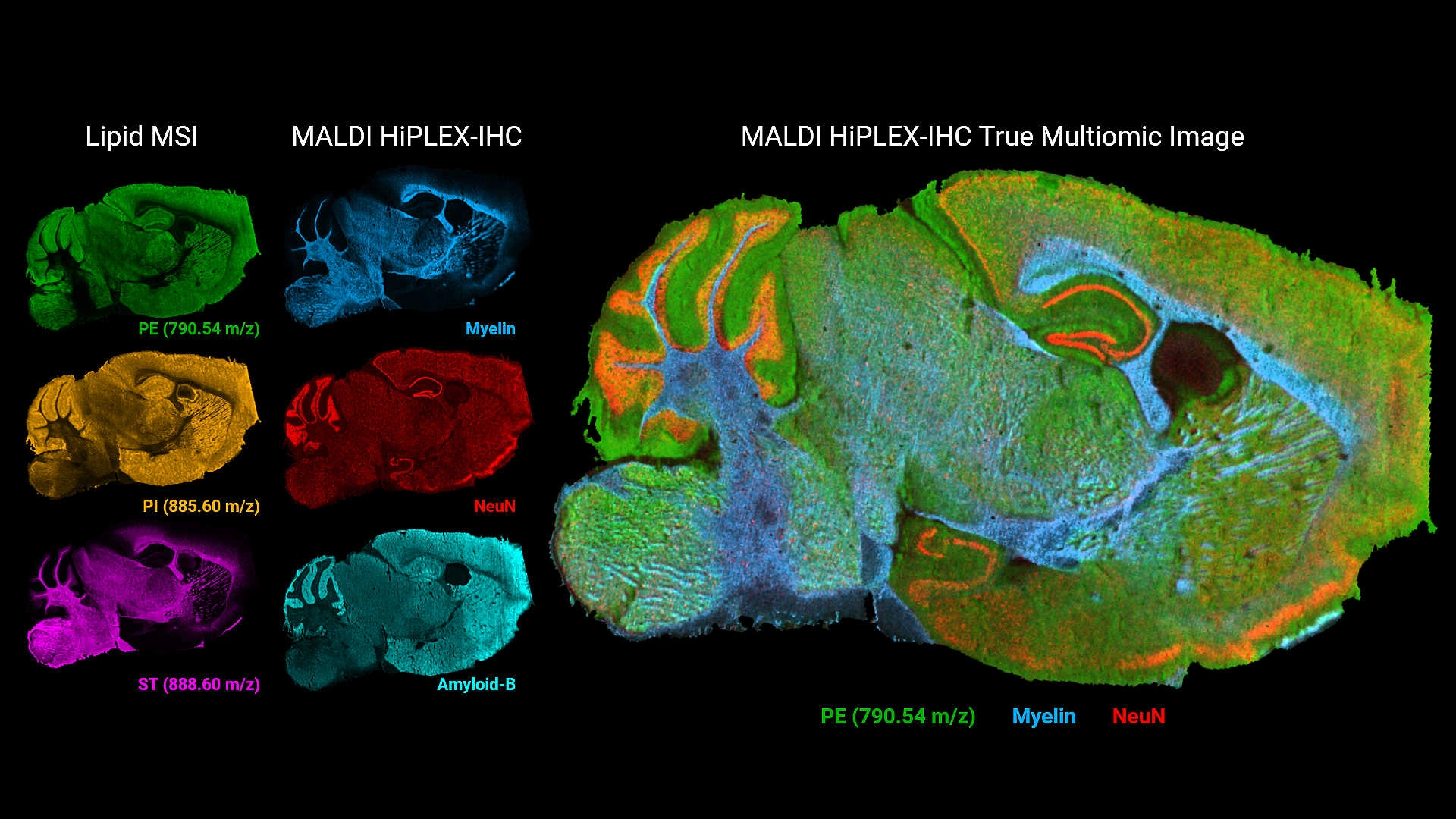
The new MALDI HiPLEX-IHC technology combines unbiased small molecule MALDI Imaging with protein profiling, enabling colocalization of glycans, lipids, and metabolites in tandem with specific proteins within tissue sections.
The strategy is enabled by AmberGen’s Miralys™ antibody-based photocleavable peptide mass tags used to map tens to over one hundred targeted proteins.
This protein imaging information is combined with MALDI Imaging data and/or immunohistochemistry (IHC) staining information of FFPE tissue samples.
The technology should provide deep, spatial, multiomic imaging of proteins and small molecules with higher levels of detail. Furthermore, this new layer of protein-specific spatial information may open a range of investigations such as tracking signaling pathways in glycosylation, mapping lipid profiles for tumor microenvironments, or observing how drugs effect both protein and metabolic states.
Image credits Bruker Daltonics.

Jan 05, 2026
Buying Guides
The Bottom Line: How Smart Microcentrifuge Tube Choices Impact Lab Efficiency and Cost
Innovative microcentrifuge tube solutions are more than just lab consumables—they’re strategic tools that can reduce costs, improve outcomes, and support long-term sustainability goals.

Jan 05, 2026
Buying Guides
Future-Proofing Your Lab with Innovative Microcentrifuge Tube Solutions
As laboratories evolve to meet the growing demands of modern science, even the most basic tools—like microcentrifuge tubes—are being reimagined.

Jan 05, 2026
Buying Guides
Microcentrifuge Tube Innovations: Driving Sustainable Change
Integrating next-generation consumables into standard workflows can lead to a more sustainable and responsible research environment.

Jan 05, 2026
Buying Guides
Single Use Plastics: How to Manage these Major Contributors to Lab Waste
Reducing lab waste isn’t just a sustainability issue—it’s a financial and operational one, too.

Jan 05, 2026
Buying Guides
Common Microcentrifuge Tube Challenges in Sensitive Molecular Workflows
Key factors that can impact the performance of microcentrifuge tubes for PCR, qPCR, and NGS and other sensitive applications

Jan 05, 2026
Buying Guides
How to Research the Right Microcentrifuge Tubes for Your Application
Selecting the right microcentrifuge tubes starts with understanding the specific demands of your application.

Jan 05, 2026
Technical Insight
The Best Confocal Microscopes of 2026
Discover the best confocal microscopes of 2026 for advanced imaging. Compare top models from Zeiss, Evident, and Nikon for speed, resolution, and budget.

Dec 24, 2025
Featured, Popular Products, Technical Insight
Emerging Mass Spectrometry Technologies to Improve Intraoperative Diagnostics
Advances in mass spectrometry may soon reshape surgery, improving diagnostic accuracy and impacting post-operative outcomes

Dec 23, 2025
Technical Insight
The Best Microplate Washers/Dispensers of 2026
Discover the top microplate washers/dispensers for 2026. Compare the latest models from Agilent, BlueCatBio, and Thermo Fisher to optimize your lab's throughput and precision today.
Dec 15, 2025
Featured, Popular Products
Automated 3D Cell Culture Workflows: Reshaping Disease Research and Drug Development
As 3D cell systems move from exploratory research into routine use across drug development and translational science, automation is becoming essential.